Your cart
There are no more items in your cart


Made in ultra-high resolution 3D printing in full color.
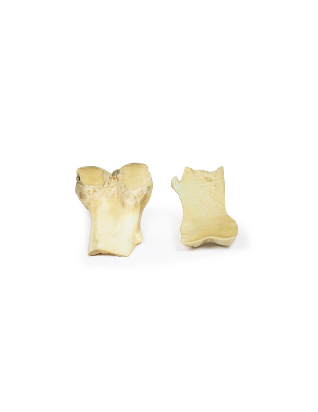
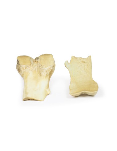
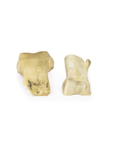
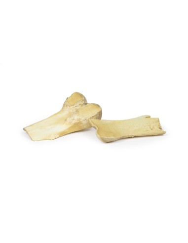
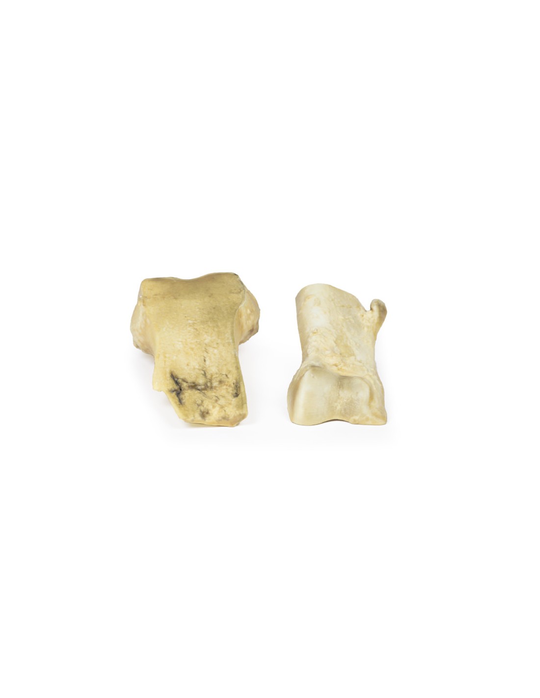
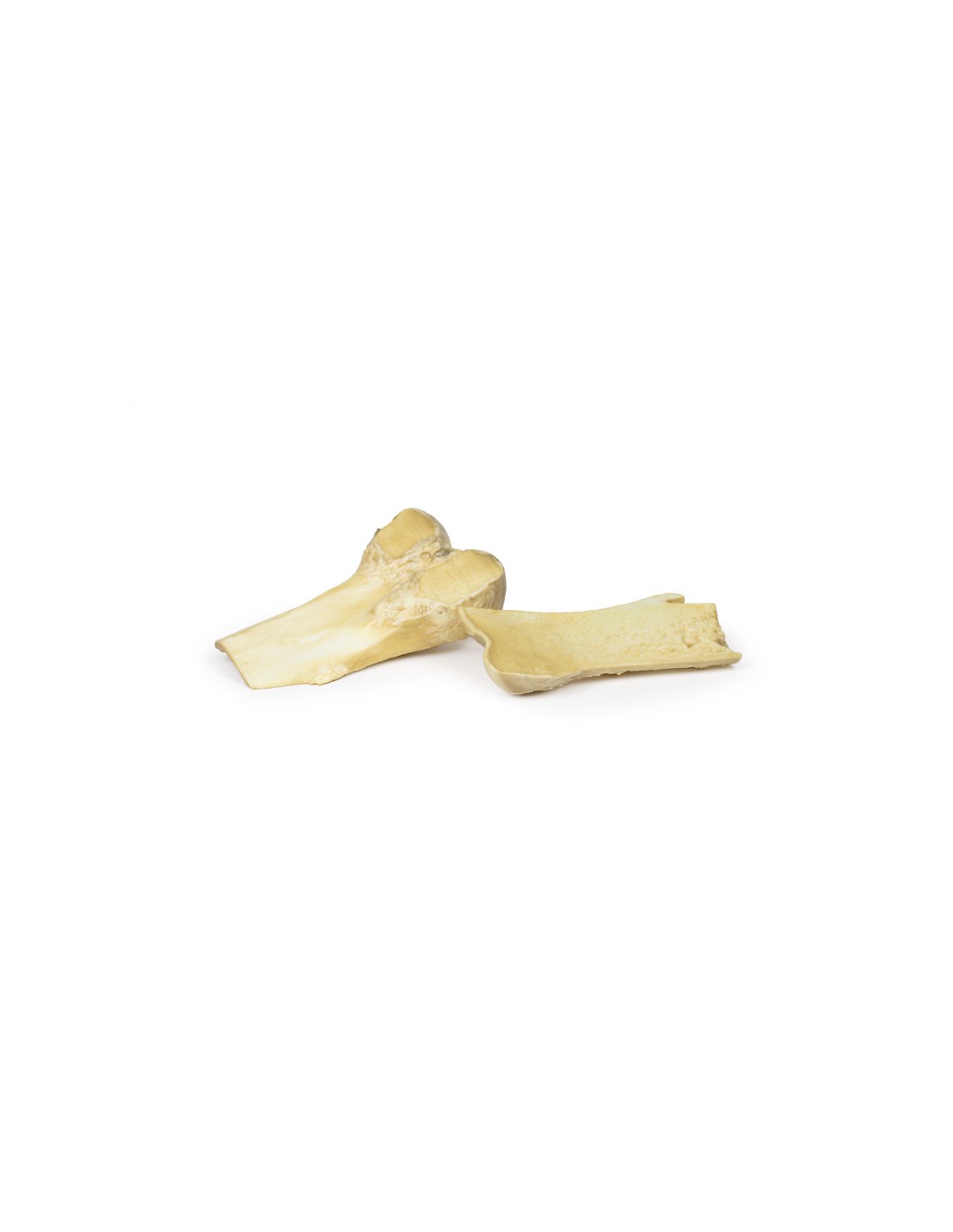
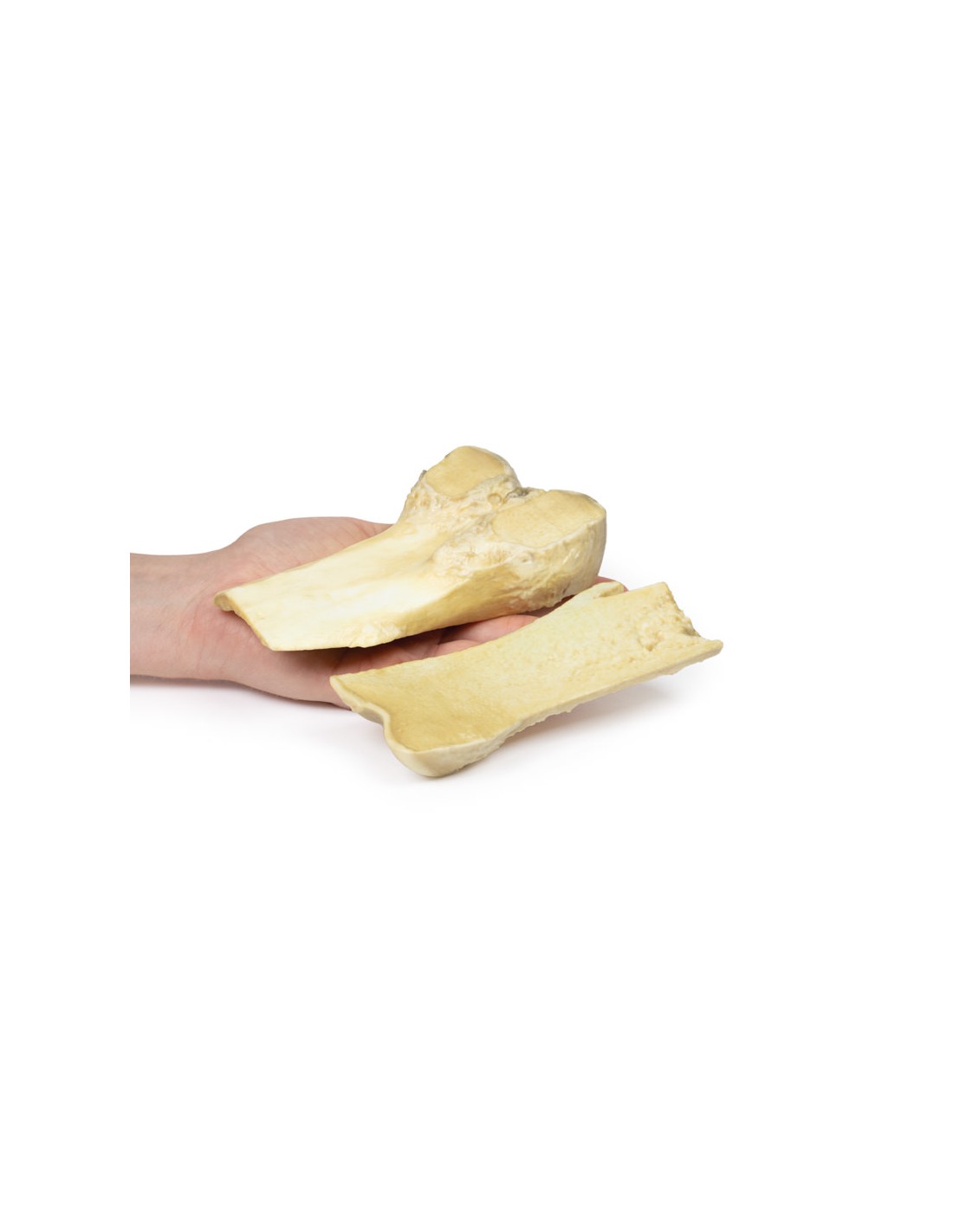
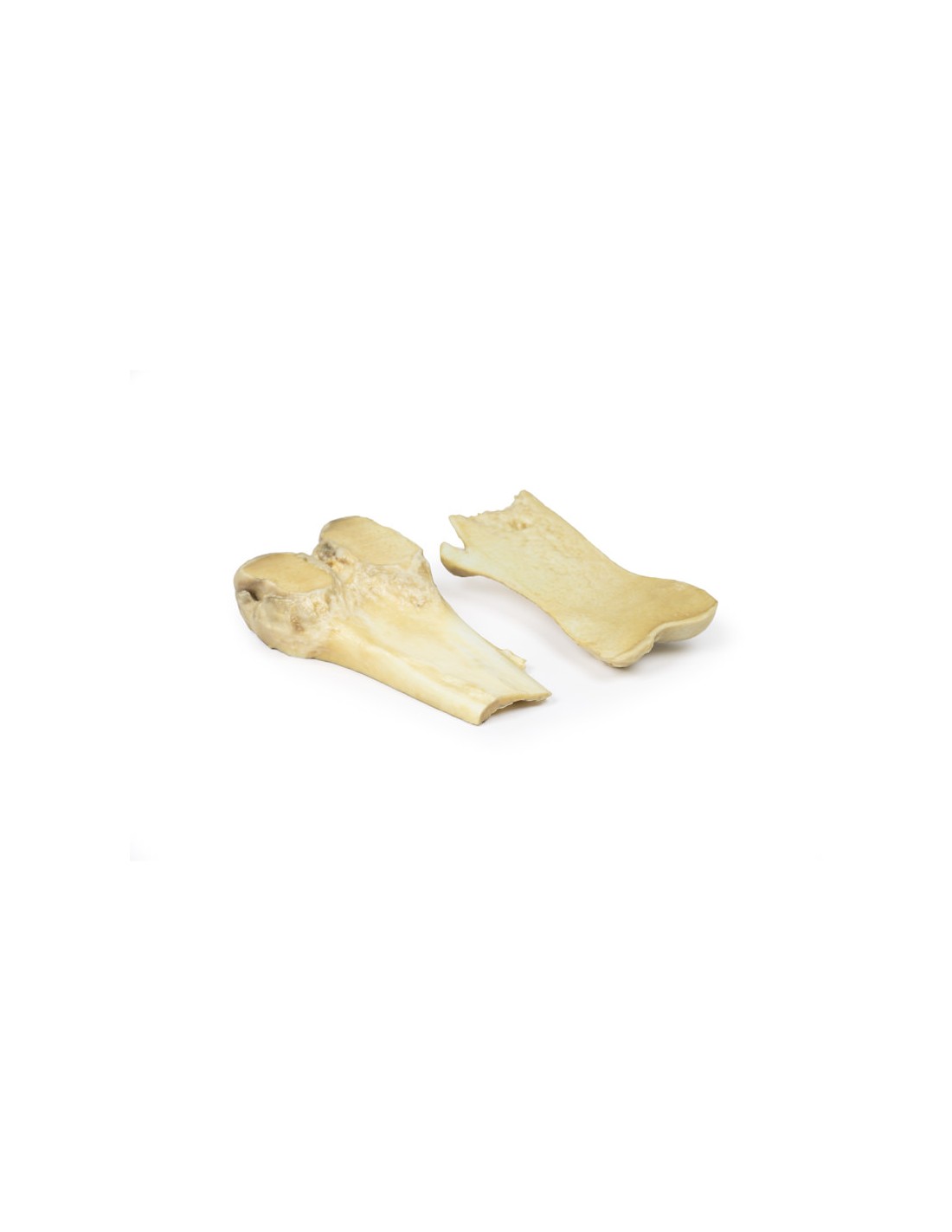
Osteochondroma - Erler Zimmer 3D anatomy Series MP2118
This dissection model highlighting an Osteochondroma is part of the exclusive Monash 3D anatomy series, a comprehensive series of human dissections reproduced with ultra-high resolution color 3D printing.
Clinical History.
A 61-year-old man with prostate cancer comes to the pre-assessment clinic before a prostatectomy. Overall, he feels well without major complaints. After examining his symptoms, it is noted that he has chronic pain in his right knee, which his family doctor called osteoarthritis. To rule out bone metastases of prostate cancer, an X-ray of the knee is prescribed, which shows a protruding pedunculated lesion from the medial aspect of the diaphysis of the right femur. His prostatectomy goes ahead but he subsequently dies from a postoperative pulmonary embolism.
Pathology
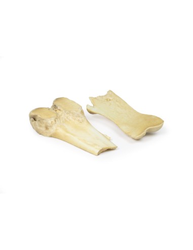
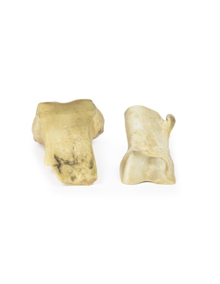
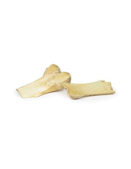
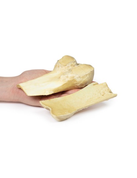
The specimen is the lower end of the patient's right femur, which was cut in the coronal plane and mounted to visualize the external surfaces. A 2-cm-long pedunculated bony protuberance protrudes from the medial aspect of the femoral diaphysis 7 cm above the medial condyle. The projection is composed of normal bone with a thin cap of hyaline cartilage at the end. This is an example of osteochondroma.
Additional information
An osteochondroma (or exostosis) is a benign cartilaginous tumor. They consist of a cartilaginous hooded bony protrusion from the outer surface of the bone from which they arise. They are the most common benign bone tumors. Most osteochondromas occur spontaneously, but they can also occur as part of multiple hereditary exostosis syndrome or after radiation therapy. They usually develop from or near the growth plate. Most commonly they arise from the appendicular skeleton, especially in the lower limb around the knee or in the upper limb at the proximal humerus. Men are more commonly affected than women.
Symptoms vary depending on the site and size of the growth. Many osteochondroma remain asymptomatic. Osteochondroma leads to symptoms from compression of surrounding neurovascular structures. They can also cause myositis pain or bone spur fracture. They usually occur in the second decade of life. They can be diagnosed with a simple radiograph, but MRI is the gold standard to ensure that there are no malignant tumors within the growth.
Hereditary exostoses are associated with mutations in the EXT1 and EXT2 genes. Reduced expression of these genes has also been observed in sporadic osteochondromas. Osteochondromas stop forming when fusion of growth cartilage occurs. Excision treatment is only if symptoms are severe. Malignant transformation to chondrosarcoma is rare in sporadic cases but more common in hereditary exostosis (5-20%).
What advantages does the Monash University anatomical dissection collection offer over plastic models or plastinated human specimens?
- Each body replica has been carefully created from selected patient X-ray data or human cadaver specimens selected by a highly trained team of anatomists at the Monash University Center for Human Anatomy Education to illustrate a range of clinically important areas of anatomy with a quality and fidelity that cannot be achieved with conventional anatomical models-this is real anatomy, not stylized anatomy.
- Each body replica has been rigorously checked by a team of highly trained anatomists at the Center for Human Anatomy Education, Monash University, to ensure the anatomical accuracy of the final product.
- The body replicas are not real human tissue and therefore not subject to any barriers of transportation, import, or use in educational facilities that do not hold an anatomy license. The Monash 3D Anatomy dissection series avoids these and other ethical issues that are raised when dealing with plastinated human remains.
No reviews
Tap to zoom