
Your cart
There are no more items in your cart


Osteosarcoma of the femur - Erler Zimmer 3D anatomy Series MP2115
erler zimmer
EZ-MP2115
€701.87
Tax included
Made in ultra-high resolution 3D printing in full color.
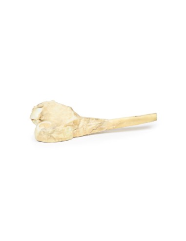
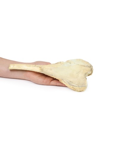
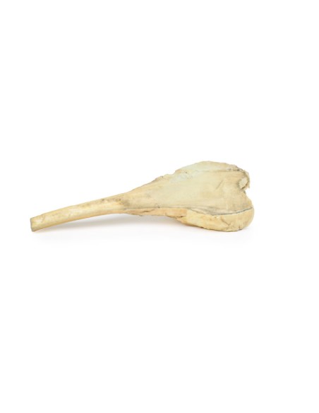
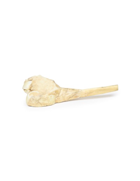
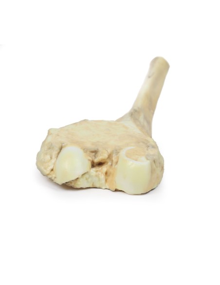
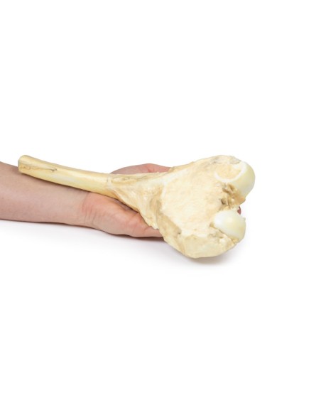
Osteosarcoma of the Femur - Erler Zimmer 3D anatomy Series MP2115
This dissection model highlighting an Osteosarcoma of the femur is part of the exclusive Monash 3D anatomy series, a comprehensive series of human dissections reproduced with ultra-high resolution color 3D printing.
Clinical History.
A 16-year-old man presented with a 3-month history of increasingly swollen and painful right knee. On examination, there was palpable tender swelling above the right knee. Blood analysis showed an increased alkaline phosphatase level. An X-ray of the knee showed periosteal reactive changes in the distal femur suspicious for a bony neoplasm. The patient then underwent CT and MRI staging of the right leg. He underwent adjuvant chemotherapy before resection of the right femur. He made a full recovery.
Pathology
The specimen is the patient's resected distal femur. On the cut surface there is a large pale infiltrating tumor, 10 cm in maximum diameter, extending through the periosteum near the articular surface. This is an osteosarcoma of the femur.
Additional Information.
Osteosarcomas are a malignant tumor of bone characterized by the production of osteoid matrix or immature bone. It is the most common primary malignant tumor of bone. Most occur in the distal femur with the tibia and humerus being the most frequently affected sites. Men are more commonly affected than women. They occur in a bimodal distribution by age, with most occurring in children and adolescents under 20 years of age and the second peak in the elderly over 60 years of age.
Secondary osteosarcomas are more common in older patients. Secondary osteosarcomas occur in patient's bones with predisposing conditions such as Paget's disease, bone infarcts, and previous irradiation. Mutations in tumor suppressors and oncogenes, such as RB, TP53 and INK4a, have been shown in osteosarcomas.
Osteosarcomas usually present with painful, enlarged masses. Pathologic fractures may also be the first presenting disorder. Constitutional symptoms are usually not present. Alkaline phosphatase and lactate dehydrogenase may be elevated in blood tests. X-rays may show features of bone destruction, a mass, or signs of a periosteal reaction, such as a radiating appearance or triangular shells of reactive bone (Codman's Triangle). MRI of the affected bone is used to assess local staging of the tumor while CT of the body is used to assess distant spread. The tumor may be biopsied in some cases.
Lungs are the most common site for distant metastasis, followed by bone and brain. Treatment involves neoadjuvant chemotherapy followed by surgery. The 5-year survival rate for localized osteosarcoma is 60-70%, but drops to <20% in patients with distant metastases.
What advantages does the Monash University anatomical dissection collection offer over plastic models or plastinated human specimens?
- Each body replica has been carefully created from selected patient X-ray data or human cadaver specimens selected by a highly trained team of anatomists at the Monash University Center for Human Anatomy Education to illustrate a range of clinically important areas of anatomy with a quality and fidelity that cannot be achieved with conventional anatomical models-this is real anatomy, not stylized anatomy.
- Each body replica has been rigorously checked by a team of highly trained anatomists at the Center for Human Anatomy Education, Monash University, to ensure the anatomical accuracy of the final product.
- The body replicas are not real human tissue and therefore not subject to any barriers of transportation, import, or use in educational facilities that do not hold an anatomy license. The Monash 3D Anatomy dissection series avoids these and other ethical issues that are raised when dealing with plastinated human remains.
No reviews
Tap to zoom
