
Your cart
There are no more items in your cart


Chondrosarcoma of the femur and ileum - Erler Zimmer 3D anatomy Series MP2112
erler zimmer
EZ-MP2112
€1,109.83
Tax included
Made in ultra-high resolution 3D printing in full color.
Chondrosarcoma of the femur and ileum - Erler Zimmer 3D anatomy Series MP2112
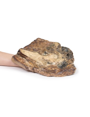
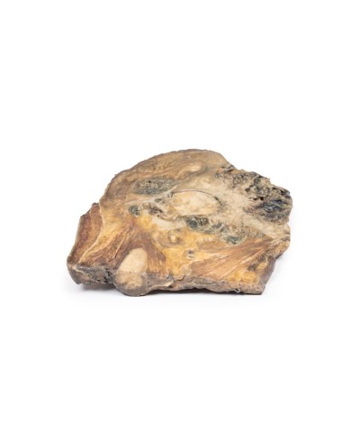
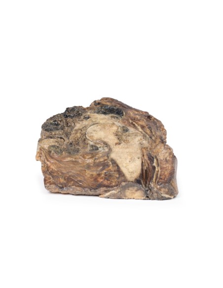
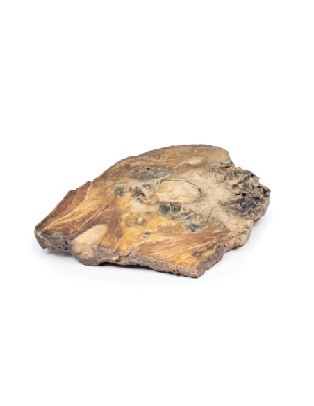
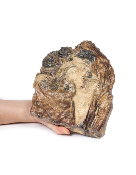
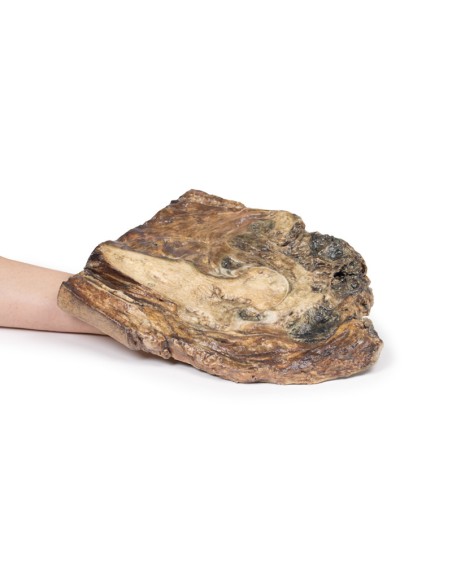
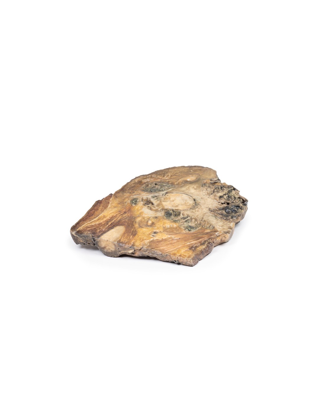
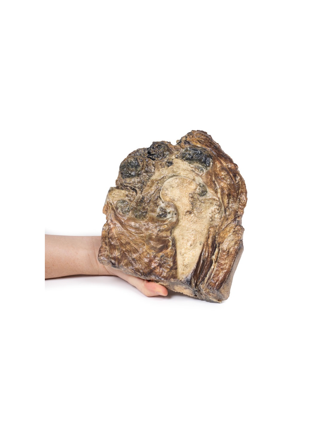
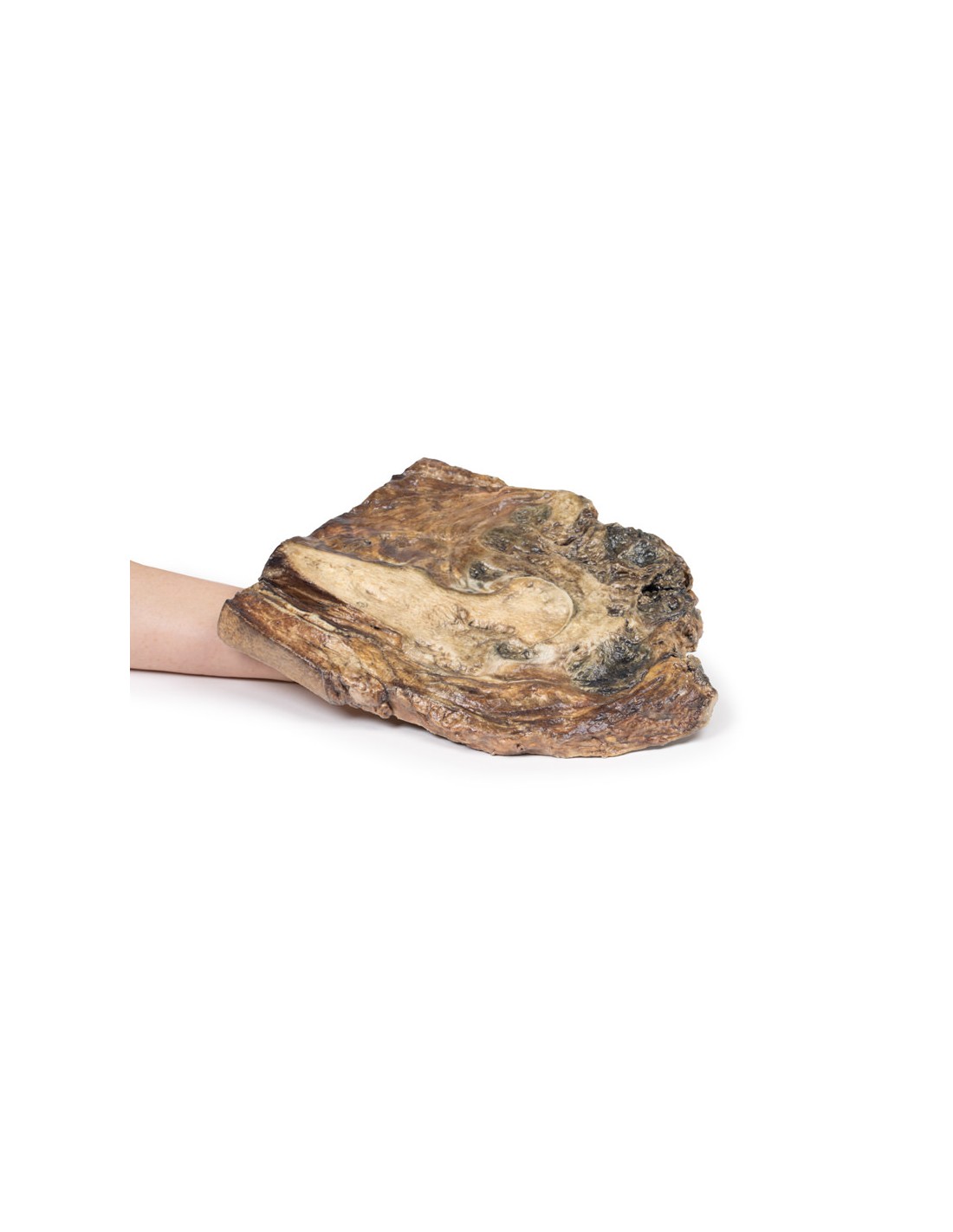
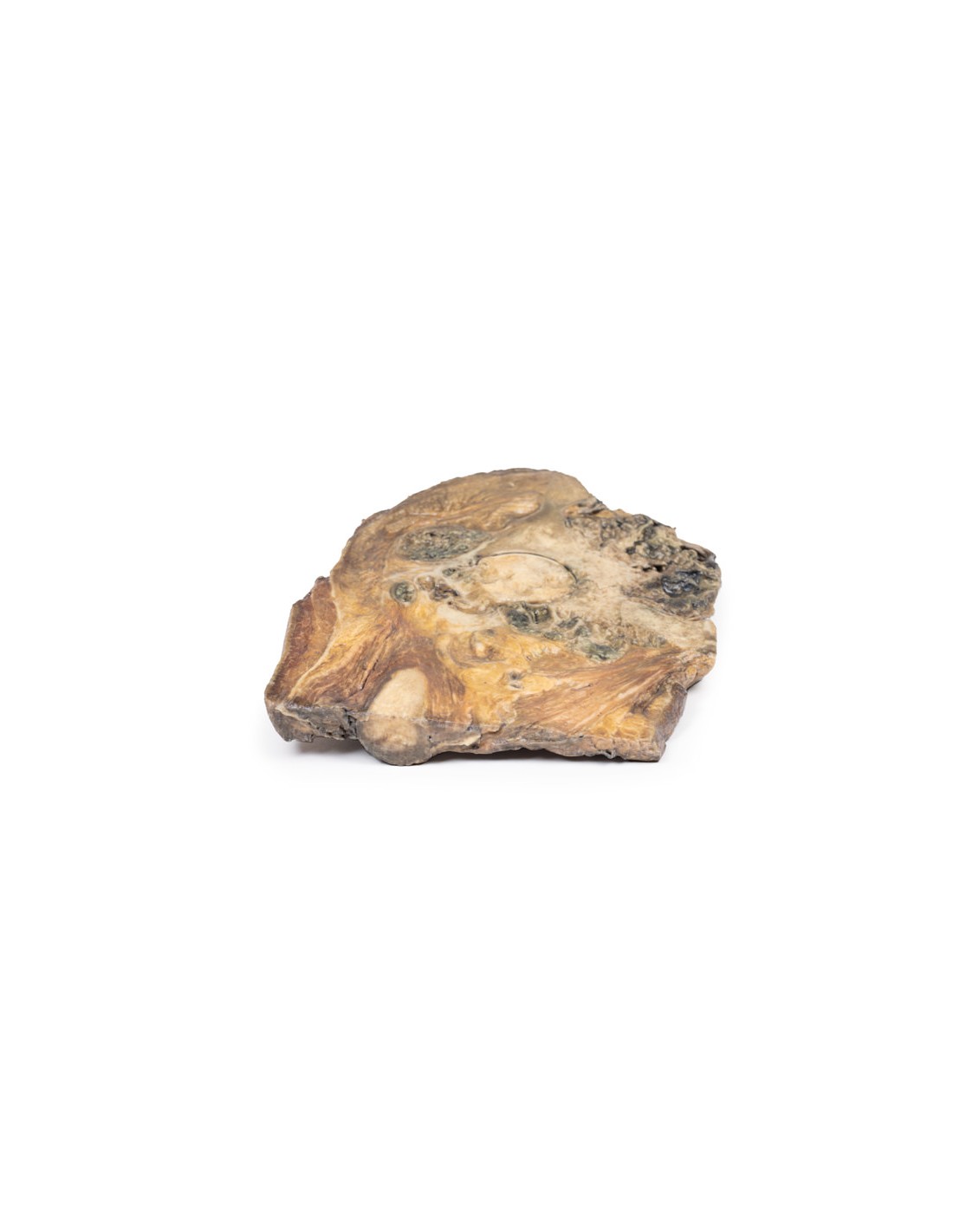
This dissection model highlighting a Chondrosarcoma of the femur and ileum is part of the exclusive Monash 3D anatomy series, a comprehensive series of human dissections reproduced with ultra-high resolution color 3D printing.
Clinical History.
A teenager presented with groin pain after horseback riding. Examination revealed a large, deep nodule. After biopsy and imaging, the diagnosis of chondrosarcoma was made and a radical surgical resection of the right leg was performed.
Pathology
The specimen consists of the upper end of the femur and its articulation with the pelvis. Within the neck and head of the femur and replacing most of the ileum is a light gray lobulated tumor with areas of cavitation, necrosis and hemorrhage. The tumor extends beyond the bone into the surrounding soft tissues and appears encapsulated. The presence of infiltration, necrosis, and hemorrhage are macroscopic features of the neoplasm.
Additional Information.
Chondrosarcoma is a primary malignant bone tumor with cartilage differentiation. It is a rare tumor that accounts for about 20% of bone tumors. The only available treatment is excisional surgical resection as current adjuvant treatments are ineffective. Pelvic location creates specific technical difficulties for both exeresis and reconstruction.
The disease usually begins in the bones of the arms, legs, or pelvis, but can be found in any part of the body that contains cartilage. Sometimes chondrosarcoma grows de novo from an otherwise healthy bone; however, sometimes it can arise from a benign bone tumor (enchondroma or osteochondroma).
There are several subtypes of chondrosarcoma, named according to their microscopic and genetic characteristics. These include: conventional chondrosarcoma; clear cell chondrosarcoma; myxoid chondrosarcoma; mesenchymal chondrosarcoma; and dedifferentiated chondrosarcoma.
What advantages does the Monash University anatomical dissection collection offer over plastic models or plastinated human specimens?
- Each body replica has been carefully created from selected patient X-ray data or human cadaver specimens selected by a highly trained team of anatomists at the Monash University Center for Human Anatomy Education to illustrate a range of clinically important areas of anatomy with a quality and fidelity that cannot be achieved with conventional anatomical models-this is real anatomy, not stylized anatomy.
- Each body replica has been rigorously checked by a team of highly trained anatomists at the Center for Human Anatomy Education, Monash University, to ensure the anatomical accuracy of the final product.
- The body replicas are not real human tissue and therefore not subject to any barriers of transportation, import, or use in educational facilities that do not hold an anatomy license. The Monash 3D Anatomy dissection series avoids these and other ethical issues that are raised when dealing with plastinated human remains.
No reviews
Tap to zoom
