
Your cart
There are no more items in your cart


Metastatic malignant melanoma - Erler Zimmer 3D anatomy Series MP2117
erler zimmer
EZ-MP2117
€476.41
Tax included
Made in ultra-high resolution 3D printing in full color.
Metastatic Malignant Melanoma - Erler Zimmer 3D anatomy Series MP2117
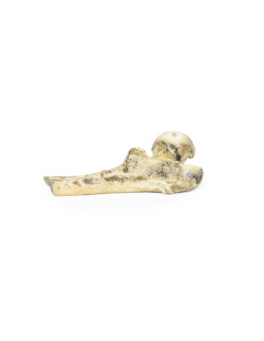
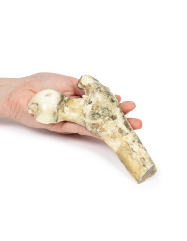
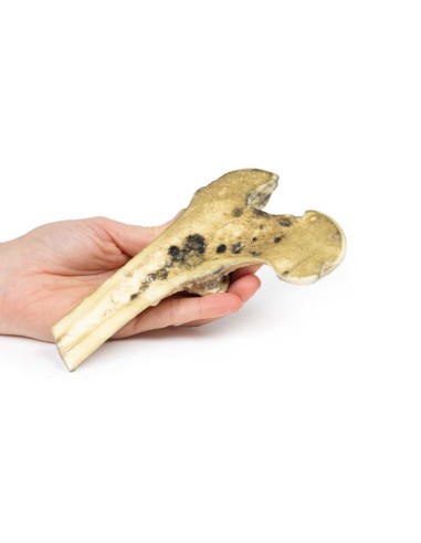
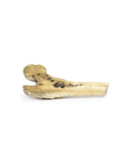
This dissection model highlighting a metastatic malignant melanoma is part of the exclusive Monash 3D anatomy series, a comprehensive series of human dissections reproduced with ultra-high resolution color 3D printing.
Clinical History.
A 65-year-old man with left groin pain. He has a history of cutaneous melanoma of the left foot treated with surgical resection and radiation therapy. On examination, he is cachexic with a hard, enlarged liver and has a discharge sinus in the left groin surrounded by black nodules. He is hospitalized and dies of hospital-acquired pneumonia.
Pathology
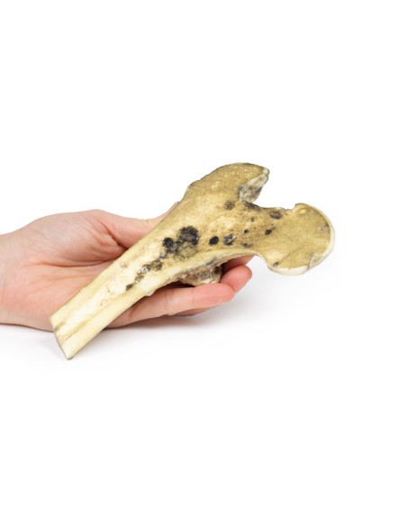
The specimen is the patient's proximal right femur sawn longitudinally to visualize the cut surface. The medullary cavity contains many deposits of tumor tissue ranging in color from pale brown to black. The spongy bone has been completely destroyed by the larger deposits, which appear dark and measure up to 3 cm in maximum diameter. Elsewhere the light brown tumor diffusely infiltrates the marrow cavity. Cortical bone has been spared, although at the junction of the shaft and neck, medially the cortical bone is discolored and thickened. These are metastatic deposits from skin melanoma.
Further information
Melanoma is a malignant skin cancer associated with UV exposure to sunlight or tanning beds. Other risk factors for developing melanoma include fair complexion, presence of a large number of melanocytic nevi, severe sunburn as a child, and immunosuppression. It accounts for about 5 percent of all skin cancer diagnoses but has the highest mortality rate of all skin cancers. Melanomas typically occur in sun-exposed areas as a pigmented lesion with irregular borders, variegated color, asymmetrical shape, and evolving over time. There are multiple mutations common in melanoma. Loss of the cell cycle control gene by mutation in the CDKN2A gene. Mutations in pro-growth signaling pathways such as BRAF and PI3K mutations are frequently observed in melanomas, as well as mutations that activate telomerase such as the TERT gene.
The most common sites of metastasis in malignant melanoma are the lungs, liver, brain and bones, as well as regional lymph nodes. Bone metastases are found in 25-50% of metastatic melanoma. The axial skeleton is most frequently affected by the spread of metastatic melanoma. These metastatic deposits cause pain and even pathological fractures. The likelihood of metastatic spread depends on the stage of the primary tumor, which is based on tumor depth, mitotic activity and skin ulceration, as well as lymph node and solid organ involvement.
The diagnosis of melanoma is made by excisional biopsy. Investigation for bone metastases is performed using blood tests (increased alkaline phosphatase, calcium, and LDH) and radiological investigations most commonly X-rays and CT scans, but MRI and PET can also be used. Treatment depends on the stage or tumor as well as the immune profile of the melanoma. Treatment usually involves surgical resection, chemotherapy, immunotherapy, radiation therapy, or most commonly a combination of treatments.
What advantages does the Monash University anatomical dissection collection offer over plastic models or plastinated human specimens?
- Each body replica has been carefully created from selected patient X-ray data or human cadaver specimens selected by a highly trained team of anatomists at the Monash University Center for Human Anatomy Education to illustrate a range of clinically important areas of anatomy with a quality and fidelity that cannot be achieved with conventional anatomical models-this is real anatomy, not stylized anatomy.
- Each body replica has been rigorously checked by a team of highly trained anatomists at the Center for Human Anatomy Education, Monash University, to ensure the anatomical accuracy of the final product.
- The body replicas are not real human tissue and therefore not subject to any barriers of transportation, import, or use in educational facilities that do not hold an anatomy license. The Monash 3D Anatomy dissection series avoids these and other ethical issues that are raised when dealing with plastinated human remains.
No reviews
Tap to zoom
