Your cart
There are no more items in your cart


Hepatocellular carcinoma - Erler Zimmer 3D anatomy Series MP2085
erler zimmer
EZ-MP2085
€503.25
Tax included
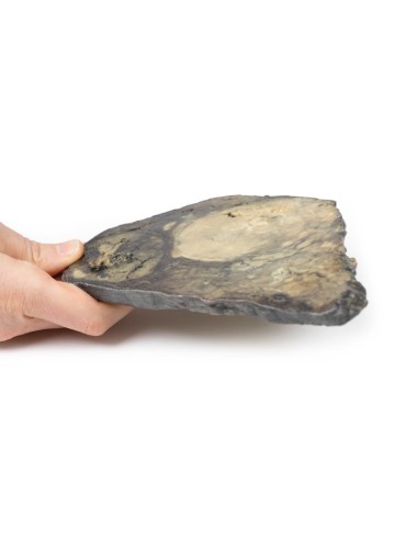
Made in ultra-high resolution 3D printing in full color.
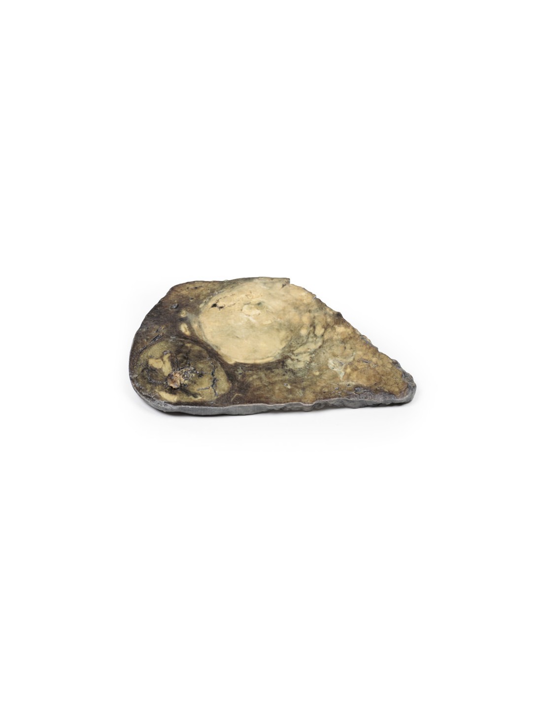
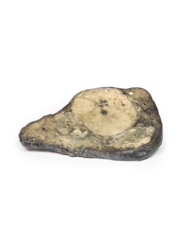
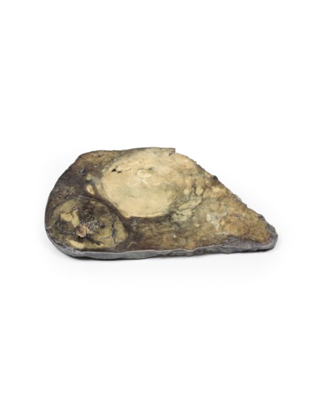
Hepatocellular Carcinoma - Erler Zimmer 3D anatomy Series MP2085
This dissection model highlighting Hepatocellular Carcinoma is part of the exclusive Monash 3D anatomy series, a comprehensive series of human dissections reproduced with very high resolution color 3D printing.
Clinical history.
A 60-year-old man is admitted with jaundice, melena and abdominal distension. He has a past medical history of untreated hepatitis C infection from previous intravenous drug use. Further questioning revealed a 9-month history of significant fatigue, weight loss, nausea, and intermittent right upper quadrant dull pain. Hepatic ultrasonography showed two large lesions within the liver. Immediately after admission, the patient died of suspected hemorrhage from esophageal varices.
Pathology
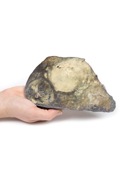
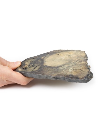
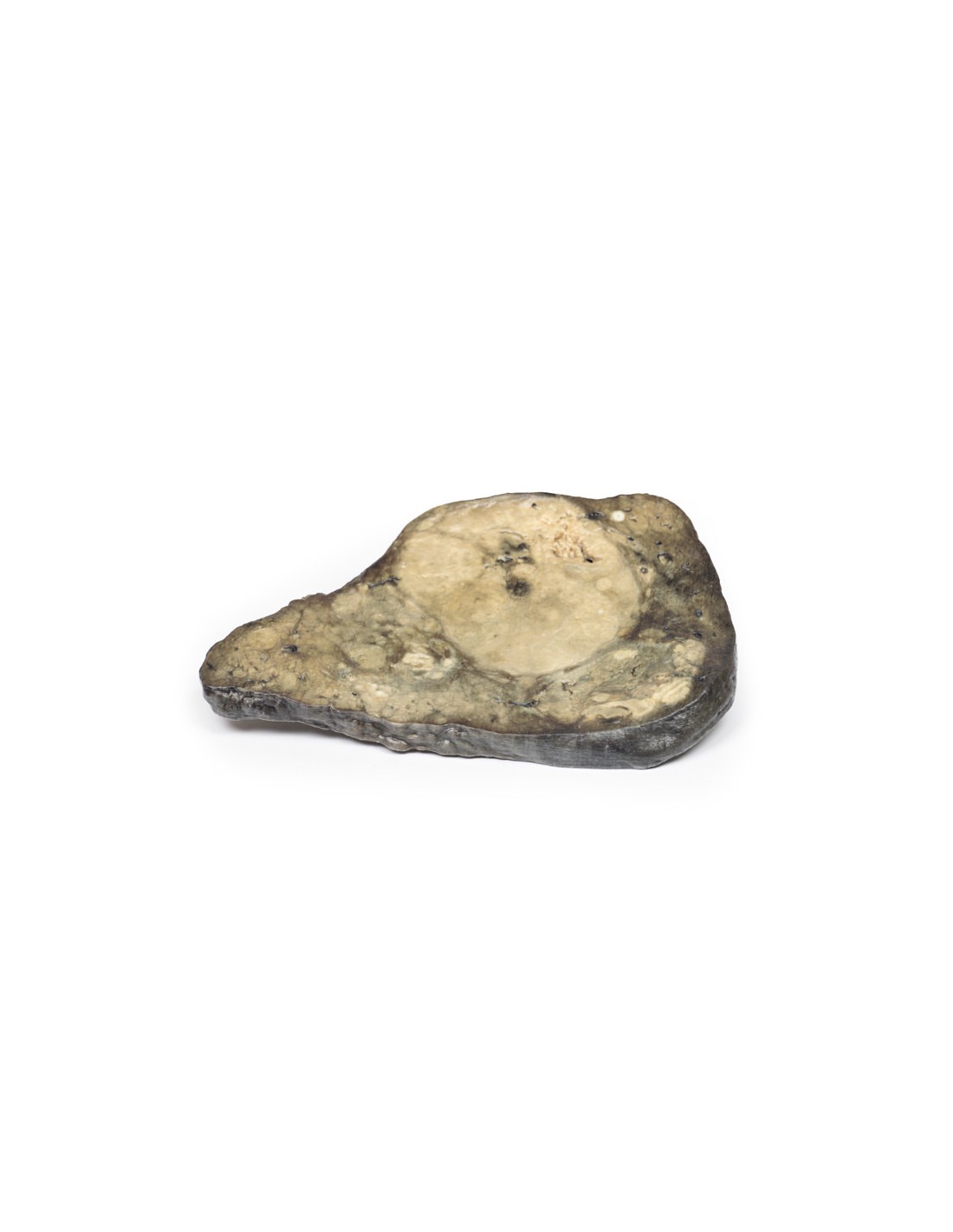
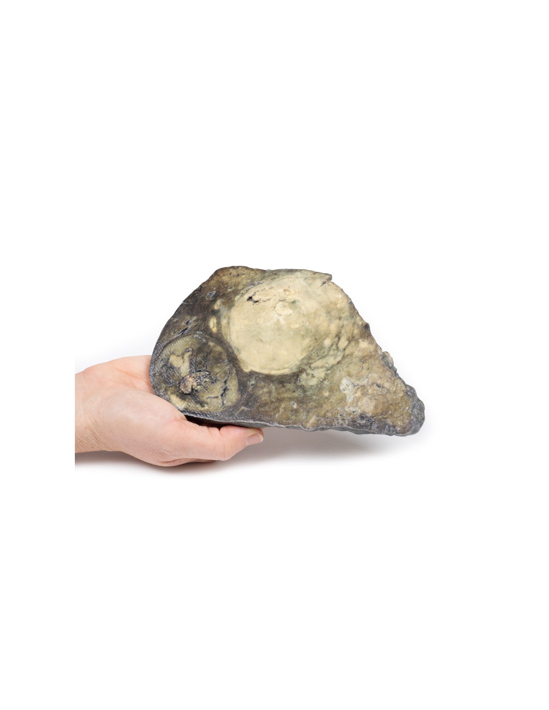
This is the patient's liver specimen at postmortem examination. The cut surface of the liver has a multinodular appearance consistent with macronodular cirrhosis. These multiple nodules vary in size up to 2 cm in diameter and are separated by narrow bands of fibrous tissue. Two large round tumors are also visible. These are 8 cm and 6 cm in diameter with a variegated cut surface due to focal necrosis, hemorrhage, and bile staining. This is an example of hepatocellular carcinoma that has developed on the background of a cirrhotic liver.
Further information
Hepatocellular carcinoma is the most common primary malignant liver tumor. HCC arises from hepatocytes in the liver. Risk factors for the development of HCC include viral infections (hepatitis B and hepatitis C), cirrhosis of the liver, aflatoxin exposure, nonalcoholic hepatic steatosis (NAFLD), hemochromatosis, and Wilson's disease. The latter is an intrinsic disorder in which excessive amounts of copper accumulate in the body, particularly in the liver, brain, and eyes. The incidence of HCC is highest in Asia and sub-Saharan Africa. There is a higher risk of developing HCC in males. HCC is associated with acquired driver mutation in oncogenes and oncosuppressor genes. The two most common driver mutations that can lead to HCC are gain-of-function mutations in beta-catenin and loss-of-function mutations in p53.
Clinically, HCC may present with abdominal pain, fatigue, weight loss, abdominal fullness and, less commonly, jaundice, gastrointestinal bleeding or bleeding from varices. Metastatic spread of HCC is hematogenous, and the most common extrahepatic sites are the lung, abdominal lymph nodes, and bone. Death usually occurs from cachexia, hemorrhage, or liver failure. Treatment varies according to the stage of the tumor and the patient's general status and underlying co-morbidities. Treatment may include surgical resection of tumor ablation, chemotherapy, and liver transplantation may be curative
.
What advantages does the Monash University anatomical dissection collection offer over plastic models or plastinated human specimens?
- Each body replica has been carefully created from selected patient X-ray data or human cadaver specimens selected by a highly trained team of anatomists at the Monash University Center for Human Anatomy Education to illustrate a range of clinically important areas of anatomy with a quality and fidelity that cannot be achieved with conventional anatomical models-this is real anatomy, not stylized anatomy.
- Each body replica has been rigorously checked by a team of highly trained anatomists at the Center for Human Anatomy Education, Monash University, to ensure the anatomical accuracy of the final product.
- The body replicas are not real human tissue and therefore not subject to any barriers of transportation, import, or use in educational facilities that do not hold an anatomy license. The Monash 3D Anatomy dissection series avoids these and other ethical issues that are raised when dealing with plastinated human remains.
No reviews
Tap to zoom