Your cart
There are no more items in your cart


Ileo with calculations - Erler Zimmer 3D anatomy Series MP2078
erler zimmer
EZ-MP2078
€420.05
Tax included
Made in ultra-high resolution 3D printing in full color.
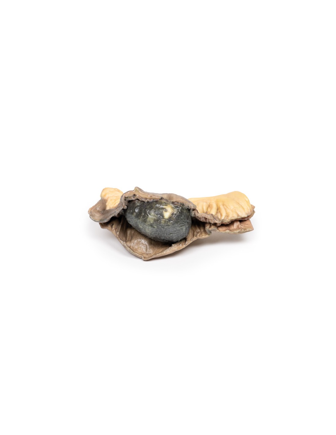
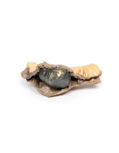
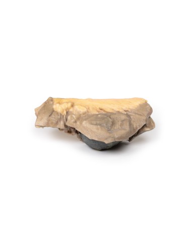
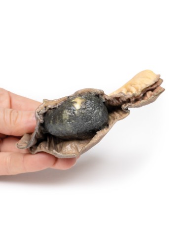
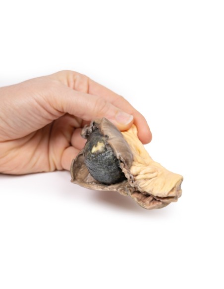
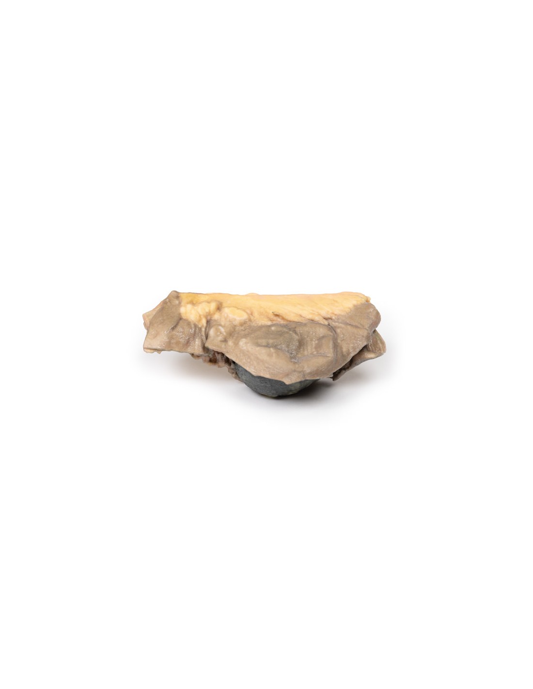
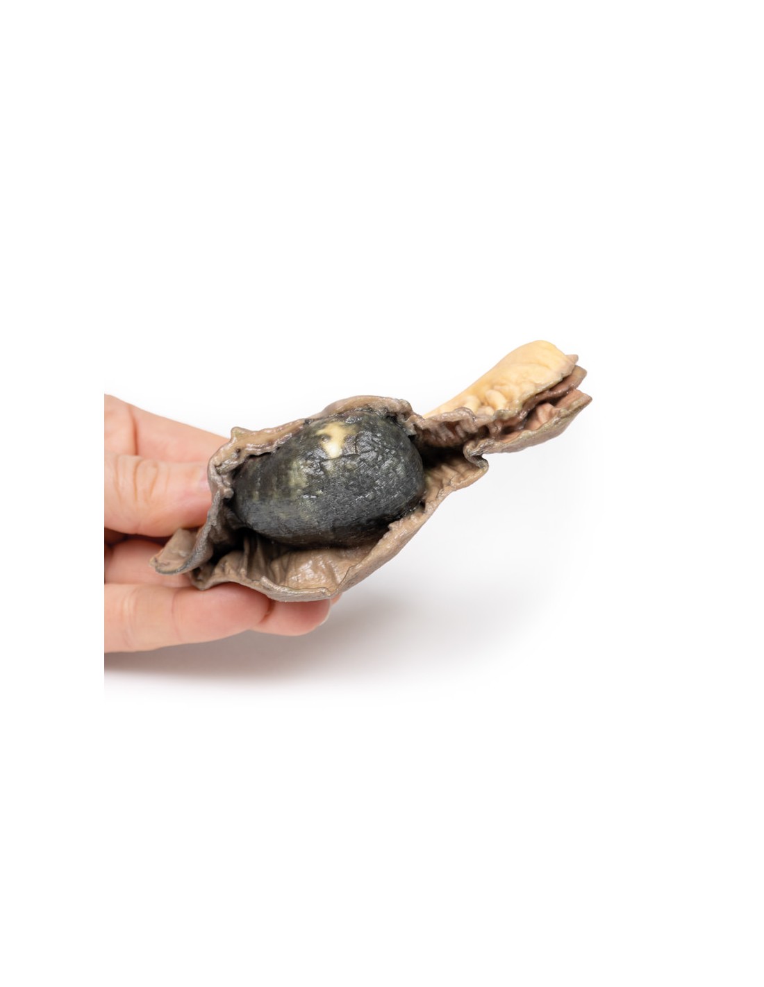
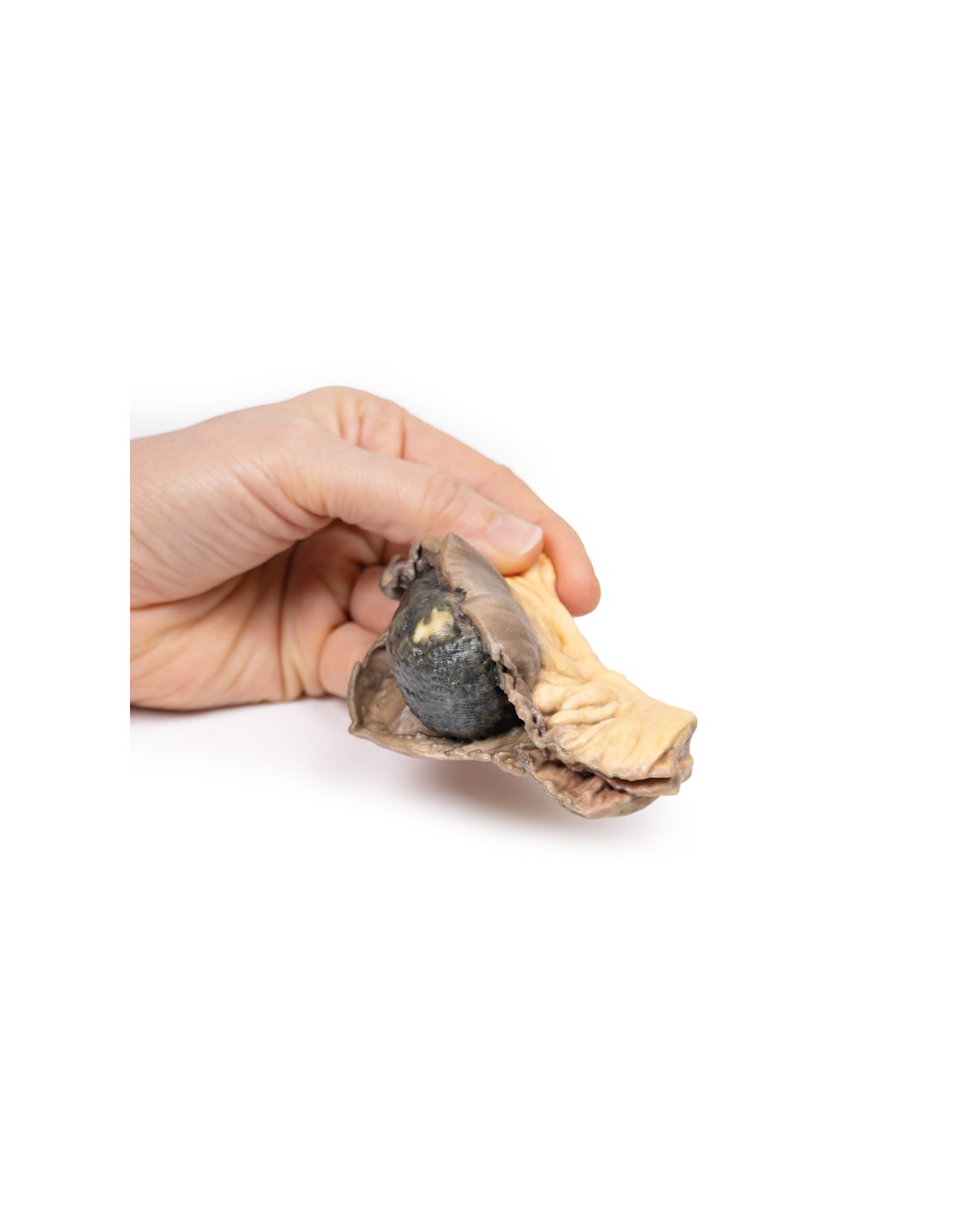
Ileus with calculi - Erler Zimmer 3D anatomy Series MP2078
This dissection model highlighting an ileum with gallstones is part of the exclusive Monash 3D anatomy series, a comprehensive series of human dissections reproduced with ultra-high resolution color 3D printing.
Clinical History.
A 54-year-old man presented to the hospital with 12 hours of severe colic, nausea and vomiting. On history, it was noted that he had a 3-year history of intermittent right subcostal pain for which he had not seen a physician. He was diagnosed with acute intestinal obstruction and a laparotomy was performed.
Pathology
This segment of the small intestine was opened to show a large ovoid pigmented gallstone with a rough surface. This is an example of gallstone ileus.
Further information
Gallstone disease is a rare cause of intestinal obstruction, accounting for only 0.5% of cases with a preponderance for elderly and female patients. It is most commonly secondary to biliary-enteric fistulas (may involve proximal or distal portions of the intestine), but can also occur after sphincterotomy. Stones are usually greater than 2-2.5 cm and impact 70% in the ileum, while others obstruct sites of stenosis/shrinkage. History may include episodic obstructive symptoms. The diagnosis is confirmed either radiologically (often on CT scan) or at the time of removal. Rigler's triad is typical of gallstone ileus and consists of: (1) small bowel obstruction, (2) a gallstone outside the gallbladder, and (3) air in the bile ducts (pneumobilia) observed on imaging and presence of gallstones on the XR plane. Treatment generally surgical with removal of obstructive stone, Fistula closure and cholecystectomy to stop recurrence. These procedures may need to be arranged.
.
What advantages does the Monash University anatomical dissection collection offer over plastic models or plastinated human specimens?
- Each body replica has been carefully created from selected patient X-ray data or human cadaver specimens selected by a highly trained team of anatomists at the Monash University Center for Human Anatomy Education to illustrate a range of clinically important areas of anatomy with a quality and fidelity that cannot be achieved with conventional anatomical models-this is real anatomy, not stylized anatomy.
- Each body replica has been rigorously checked by a team of highly trained anatomists at the Center for Human Anatomy Education, Monash University, to ensure the anatomical accuracy of the final product.
- The body replicas are not real human tissue and therefore not subject to any barriers of transportation, import, or use in educational facilities that do not hold an anatomy license. The Monash 3D Anatomy dissection series avoids these and other ethical issues that are raised when dealing with plastinated human remains.
No reviews
Tap to zoom