Your cart
There are no more items in your cart


Intussusception of the small intestine by metastatic tumor - Erler Zimmer 3D anatomy Series MP2077
erler zimmer
EZ-MP2077
€390.52
Tax included
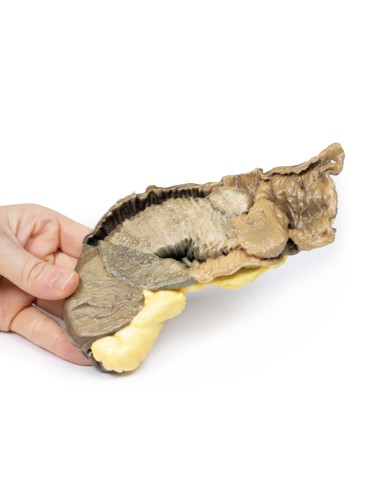
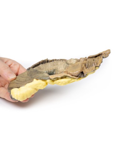
Made in ultra-high resolution 3D printing in full color.
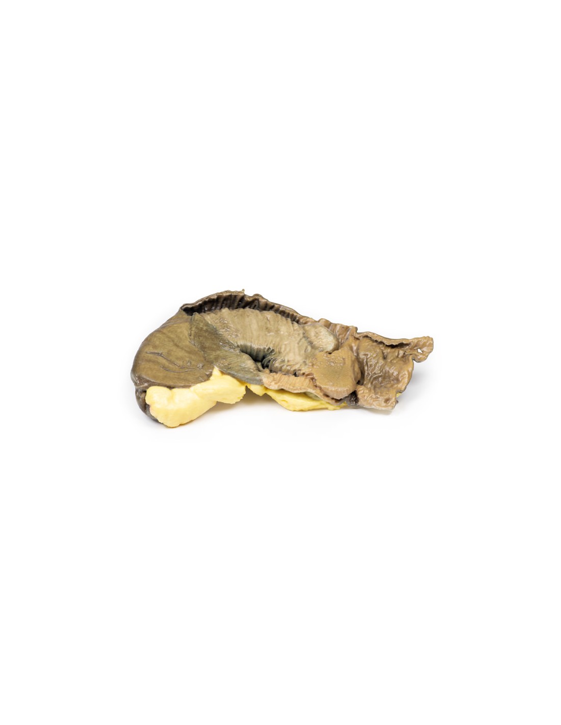
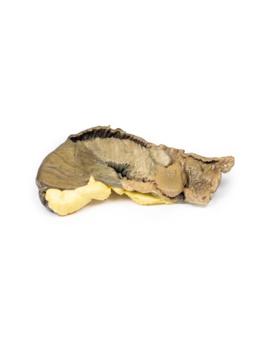
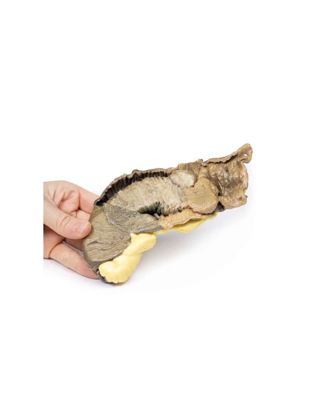
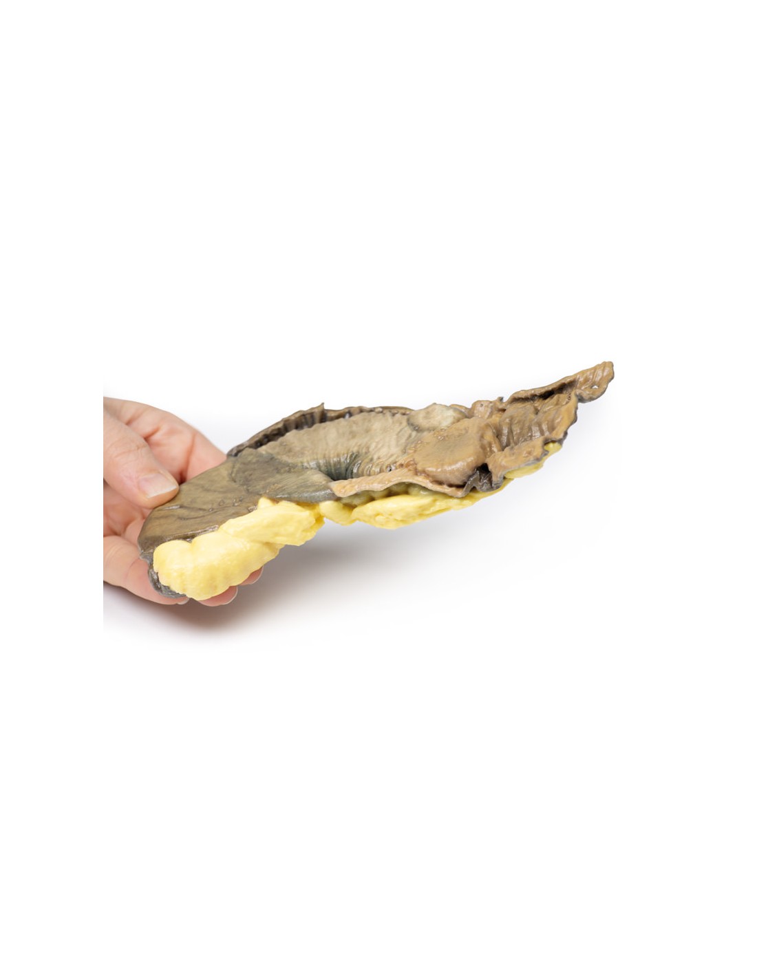
Intussusception of the small intestine from metastatic tumor - Erler Zimmer 3D anatomy Series MP2077
This dissection model highlighting an Intussusception of the small intestine from metastatic tumor is part of the exclusive Monash 3D anatomy series, a comprehensive series of human dissections reproduced with ultra-high resolution color 3D printing.
Clinical history
A 66-year-old woman suffered a sudden onset of severe central abdominal pain with colic, somewhat relieved by pulling her knees together. She passed a stool containing mucus and blood ("like currant jelly"). On examination, there was a mass in the left hypochondrium, which hardened with each spasm of pain. The specimen was excised by laparotomy.
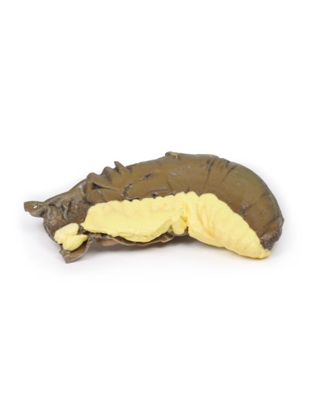
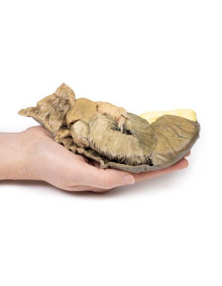
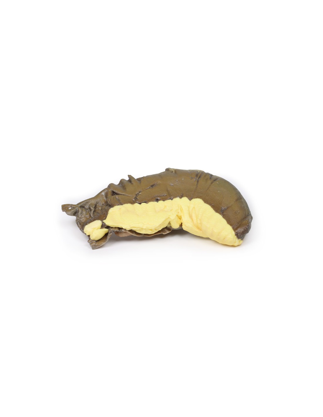
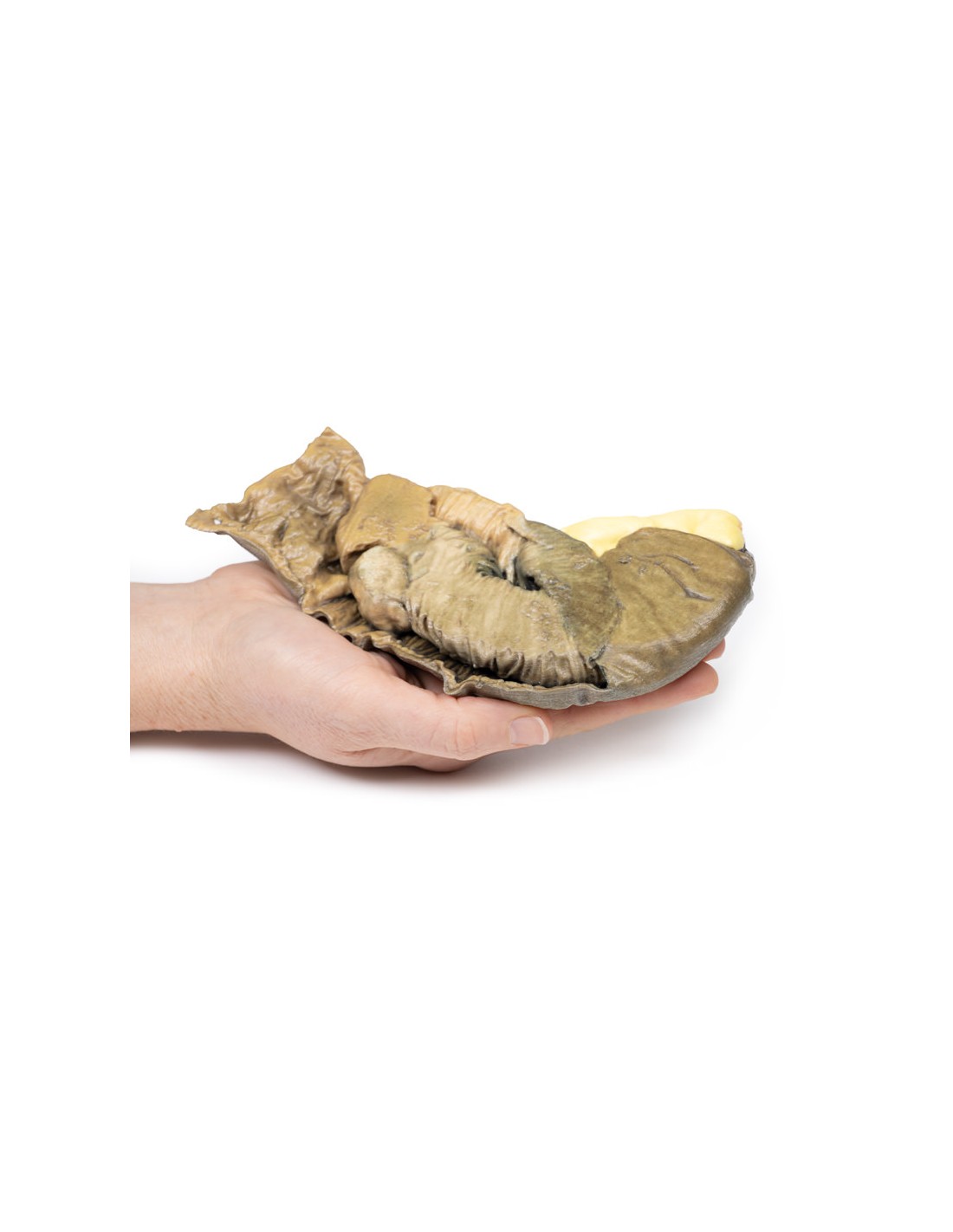
Pathology
The specimen is a segment of small intestine, about 20 cm long, with mesentery attached up to 2 cm wide (more evident on the uncut aspect of the specimen). Approximately 5 cm from the proximal surgical resection margin (located on the left side of the specimen), a 3 cm diameter polypoid tumor invaginated into the lumen of the intestine and was pushed distally, forming an invagination 13 cm in length. The tumor is visible at the apex of the intussusception (near the right side of the specimen). Congestion and exudation observed on the mucosal surface of the intussusception (invaginated portion) are features considered with early ischemic necrosis. Histologic diagnosis in this case is not recorded; however, the macroscopic appearance is consistent with a metastatic malignant tumor,
Further information
Small bowel intussusception is most common in children, in whom it is usually due to invagination of enlarged lymphoid tissue (Peyer's spots) into the wall of the distal ileum. In adults, it is rare and causes only 1 to 5 percent of cases of intestinal obstruction. The usual cause of a polypoid tumor, as seen in this specimen, acts as a pathologic lead spot that is pulled forward by peristalsis, and thus causes distal telescoping of the affected portion of the intestine. The presentation may be of intermittent symptoms of intestinal obstruction and in some cases excruciating pain. Classification of intussusception can be by causative pathology or by location. Abdominal CT scan will usually demonstrate a typical "target sign" with alternating hyper/hypodense layers.
.
What advantages does the Monash University anatomical dissection collection offer over plastic models or plastinated human specimens?
- Each body replica has been carefully created from selected patient X-ray data or human cadaver specimens selected by a highly trained team of anatomists at the Monash University Center for Human Anatomy Education to illustrate a range of clinically important areas of anatomy with a quality and fidelity that cannot be achieved with conventional anatomical models-this is real anatomy, not stylized anatomy.
- Each body replica has been rigorously checked by a team of highly trained anatomists at the Center for Human Anatomy Education, Monash University, to ensure the anatomical accuracy of the final product.
- The body replicas are not real human tissue and therefore not subject to any barriers of transportation, import, or use in educational facilities that do not hold an anatomy license. The Monash 3D Anatomy dissection series avoids these and other ethical issues that are raised when dealing with plastinated human remains.
No reviews
Tap to zoom