Your cart
There are no more items in your cart


Cholelithiasis (gallstones)- Erler Zimmer 3D anatomy Series MP2075
erler zimmer
EZ-MP2075
€335.50
Tax included
Made in ultra-high resolution 3D printing in full color.
Cholelithiasis (gallstones)- Erler Zimmer 3D anatomy Series MP2075
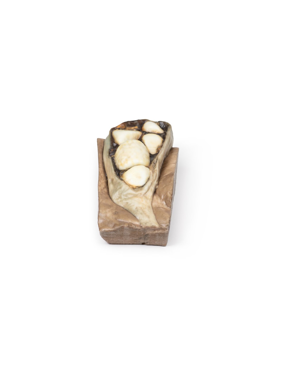
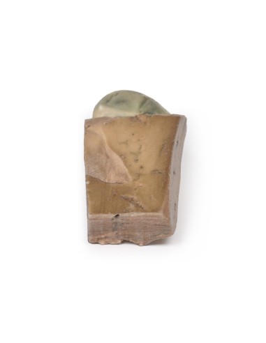
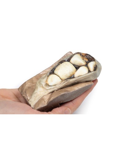
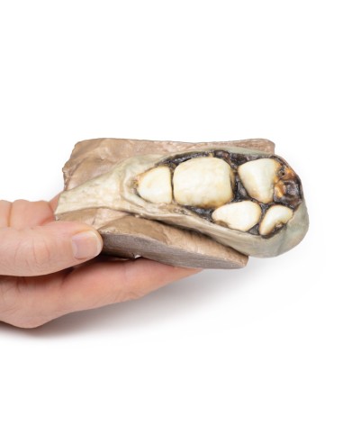
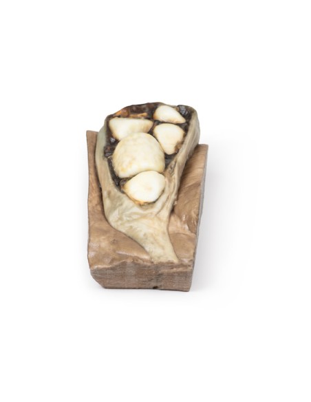
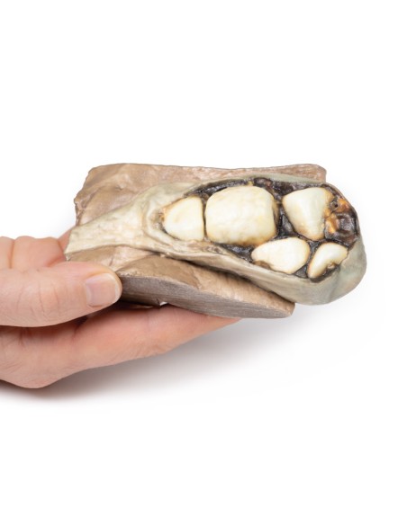
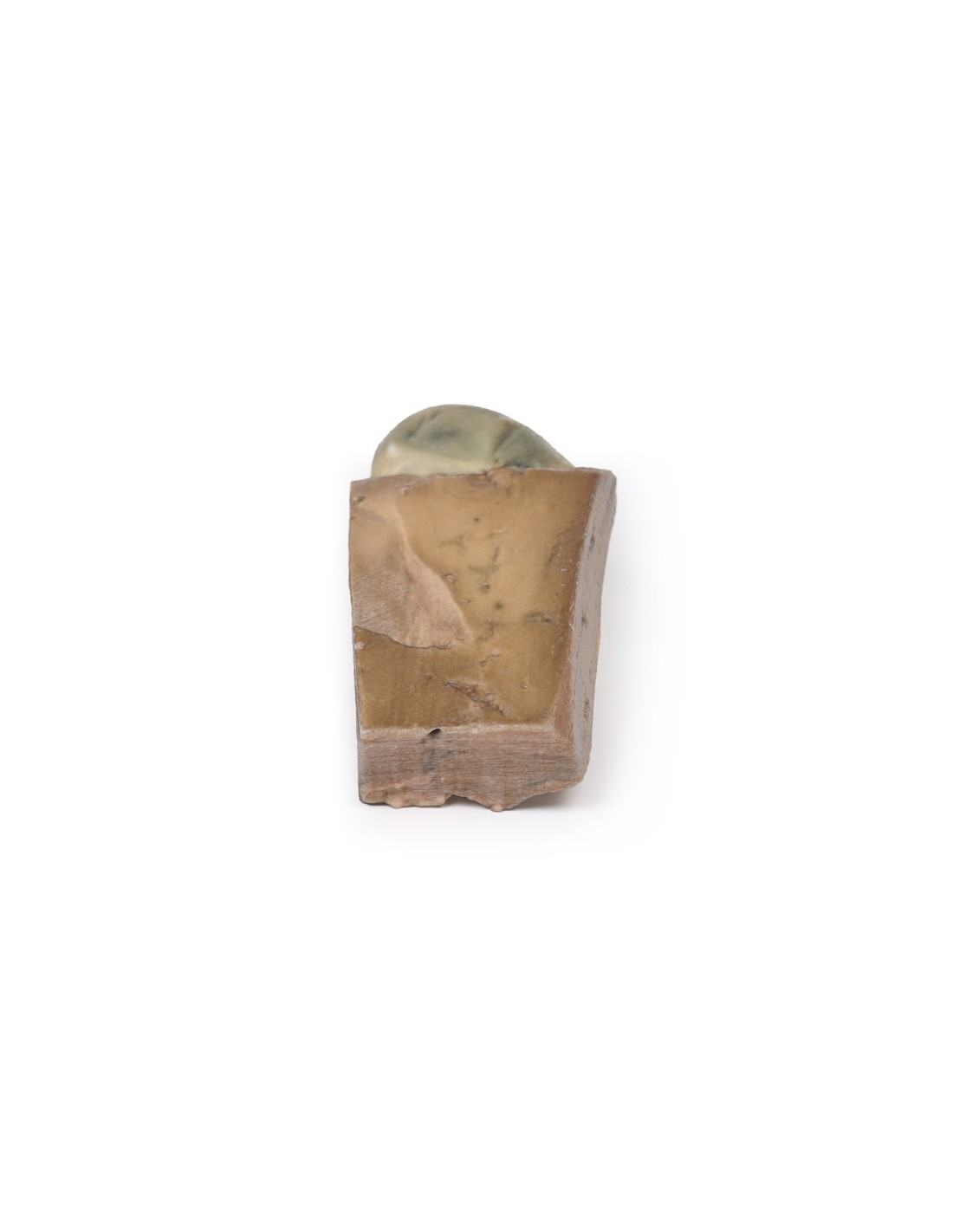
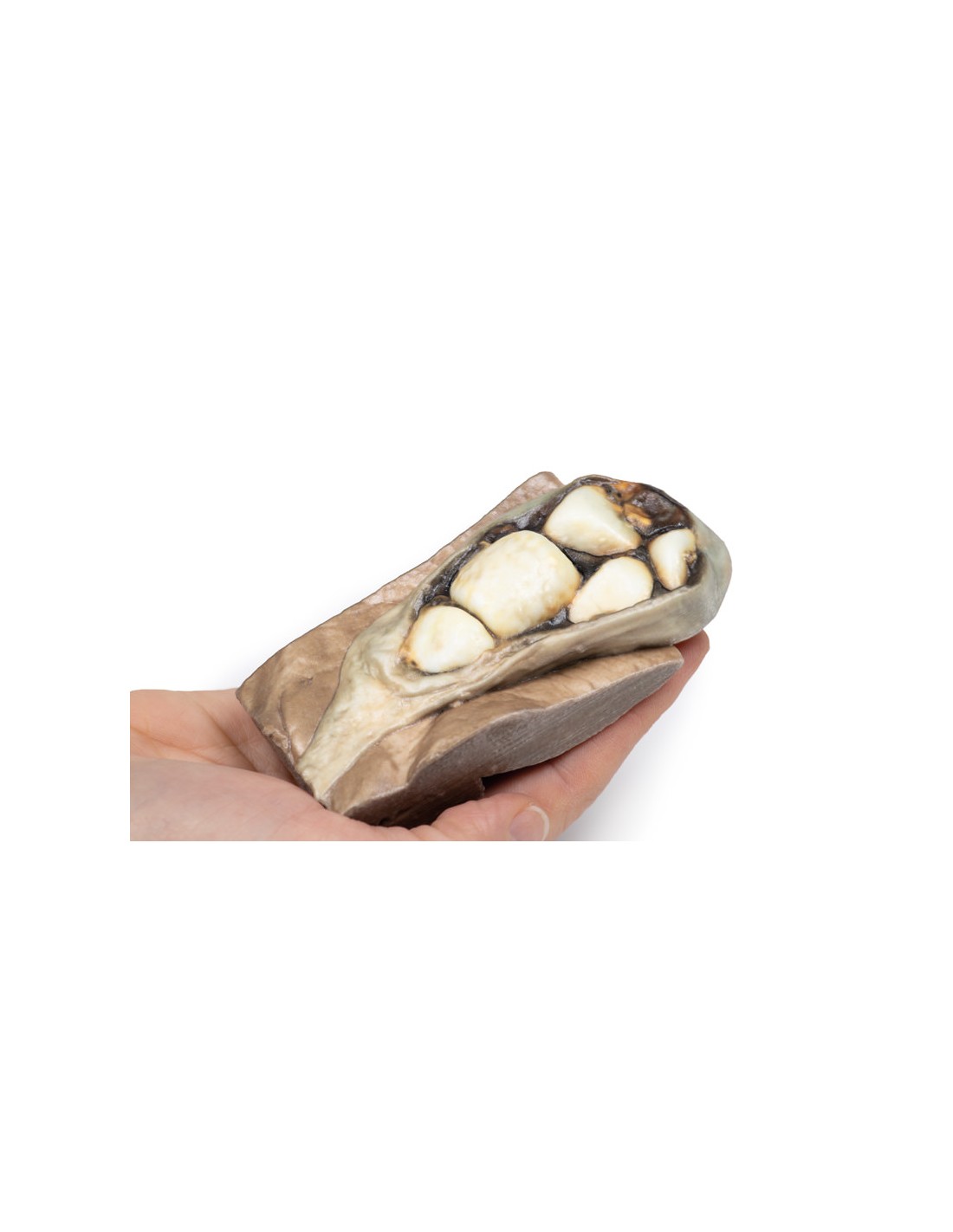
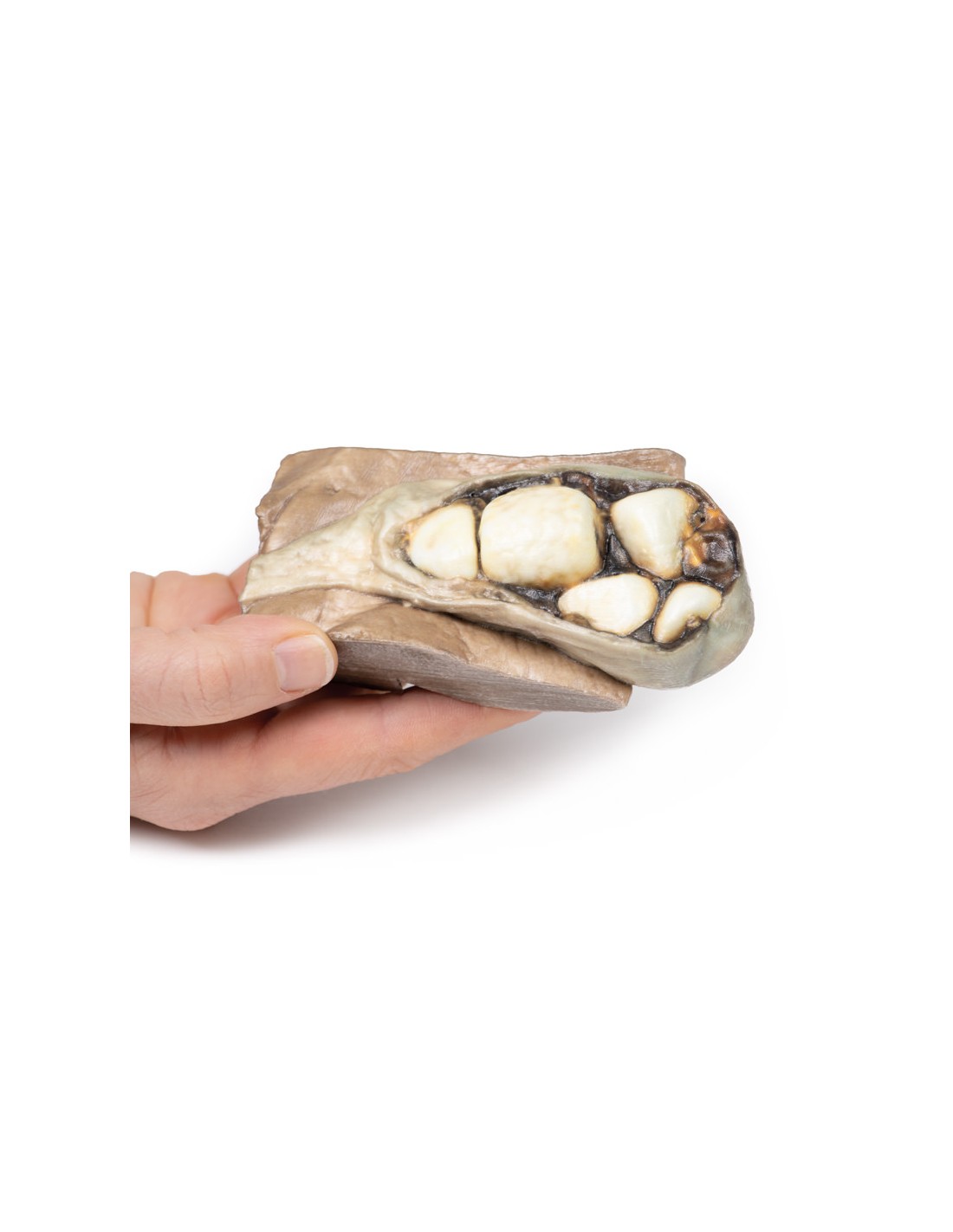
This dissection model highlighting a Cholelithiasis is part of the exclusive Monash 3D anatomy series, a comprehensive series of human dissections reproduced with ultra-high resolution color 3D printing.
Clinical History.
A middle-aged woman was studied for recurrent attacks of epigastric pain. Endoscopy revealed no peptic ulcer. A cholangiogram demonstrated a nonfunctioning gallbladder. She died of a myocardial infarction Unfortunately, the patient died at a later stage after this procedure from a myocardial infarction."
Pathology
The specimen is a portion of liver with attached gallbladder, which was opened to show six large faceted mixed stones. This is an example of cholelithiasis (gallstones).
Further information
Gallstones contain a mixture of cholesterol, calcium salts, bilirubin, protein ??and mucin. There is a high prevalence in fair-skinned populations. Risk factors include age (greater than 50 years) and female sex, along with genetic factors1, pregnancy, diabetes mellitus, and dyslipidemia. Lifestyle factors, such as rapid weight loss and certain medications (e.g., erythrocin, ampicillin, octreotide, cephalosporin), can also promote gallstone formation.
Gallstones can be asymptomatic or present with a spectrum of disease ranging from uncomplicated biliary colic to infection, cholecystitis, pancreatitis, or gallstone ileus. Typical symptoms include attacks of epigastric or right upper quadrant pain, sometimes associated with eating and often with sweating, nausea, and vomiting. The pain is usually caused by the gallbladder or biliary tract contracting forcefully against a stone, thus causing increased pressure in the gallbladder and pain. The risk of developing complications of gallstones is about 2-3% per year once biliary colic develops. Diagnosis is usually by transabdominal ultrasonography, which has largely replaced oral cholecystography studies. Cholescintigraphy (HIDA Scan) can be used to distinguish biliary colic from acute cholecystitis. Treatment of attacks is initially with simple analgesia and then definitive management usually includes elective laparoscopic cholecystectomy. Very severe cases can be life-threatening, but deaths from gallstones are rare.
It should be noted that epigastric pain can also be caused by myocardial ischemia, particularly in females, which does not necessarily present with the classic "left shoulder tip pain" of an acute myocardial infarction. Therefore, gallstones should be excluded in a patient presenting with epigastric pain, an ECG should always be performed, to help rule out heart disease[1].
Scientific references
1. Portincasa P; Moscheta A; Palasciano G (2006) Cholesterol gallstone disease. Lancet. 2006; 368(9531):230-9
What advantages does the Monash University anatomical dissection collection offer over plastic models or plastinated human specimens?
- Each body replica has been carefully created from selected patient X-ray data or human cadaver specimens selected by a highly trained team of anatomists at the Monash University Center for Human Anatomy Education to illustrate a range of clinically important areas of anatomy with a quality and fidelity that cannot be achieved with conventional anatomical models-this is real anatomy, not stylized anatomy.
- Each body replica has been rigorously checked by a team of highly trained anatomists at the Center for Human Anatomy Education, Monash University, to ensure the anatomical accuracy of the final product.
- The body replicas are not real human tissue and therefore not subject to any barriers of transportation, import, or use in educational facilities that do not hold an anatomy license. The Monash 3D Anatomy dissection series avoids these and other ethical issues that are raised when dealing with plastinated human remains.
No reviews
Tap to zoom