Your cart
There are no more items in your cart


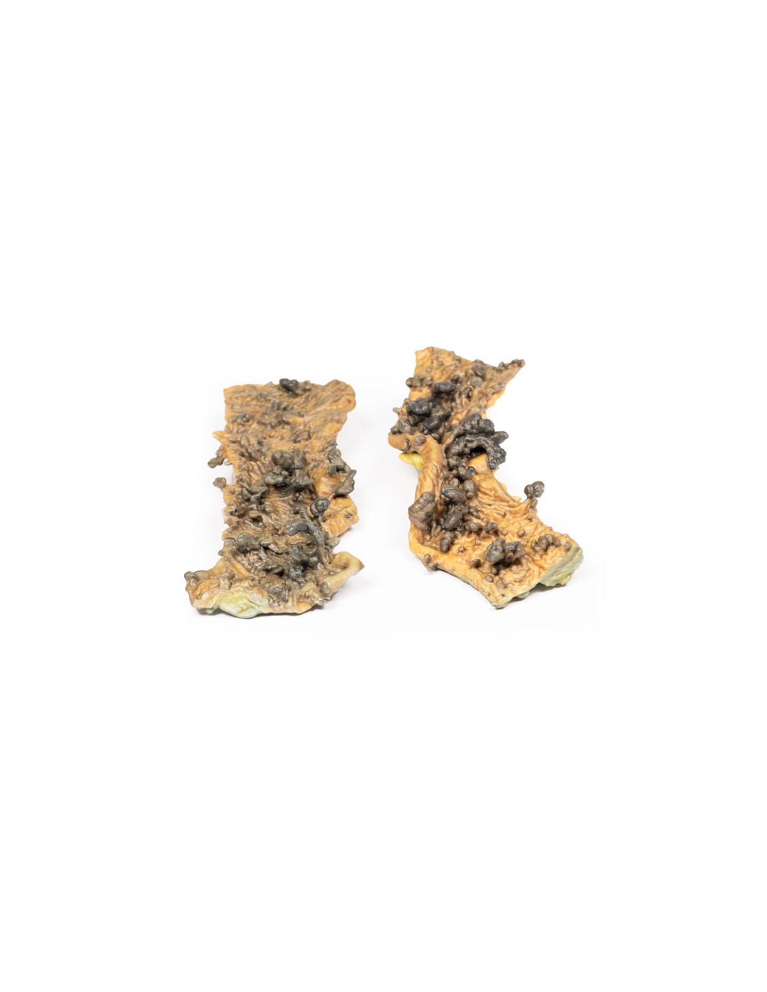
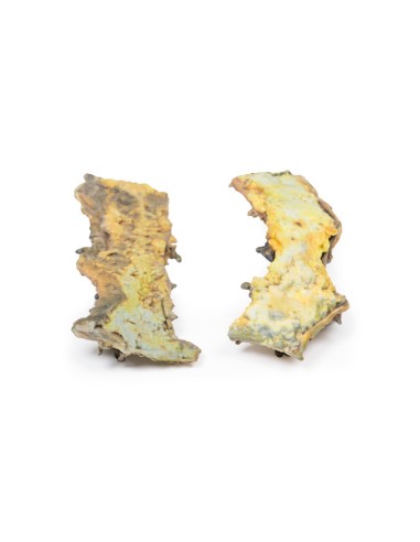
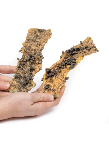
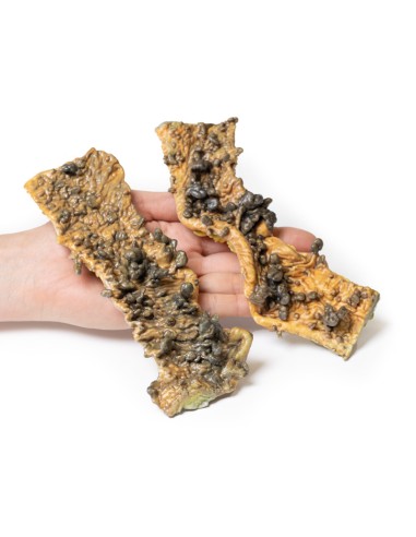
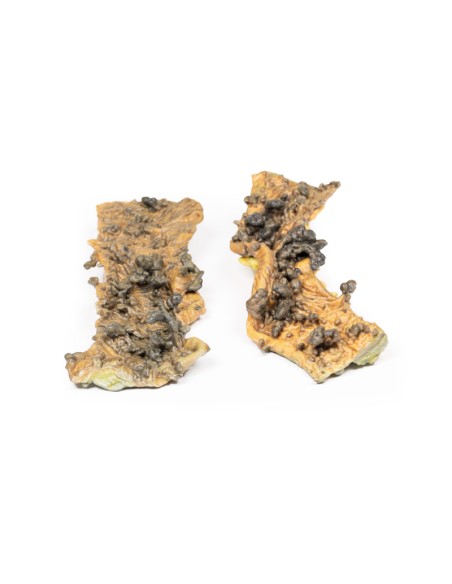
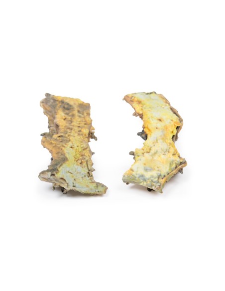
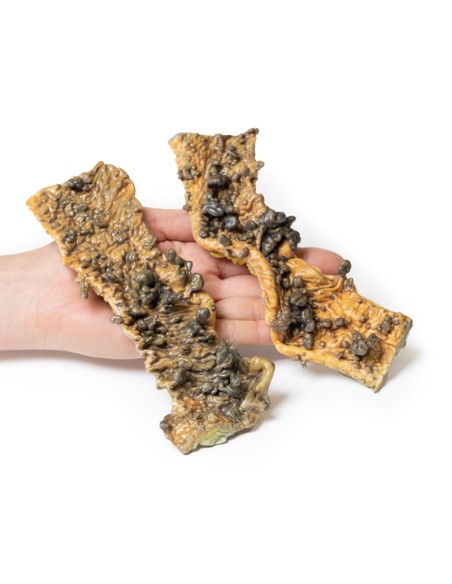
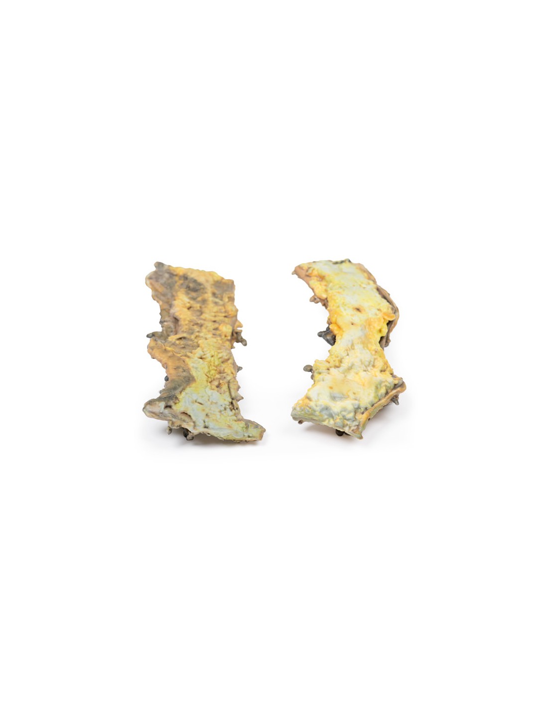
Multiple polyposis - Erler Zimmer 3D anatomy Series MP2070
erler zimmer
EZ-MP2070
€516.67
Tax included
Made in ultra-high resolution 3D printing in full color.
Multiple Polyposis - Erler Zimmer 3D anatomy Series MP2070
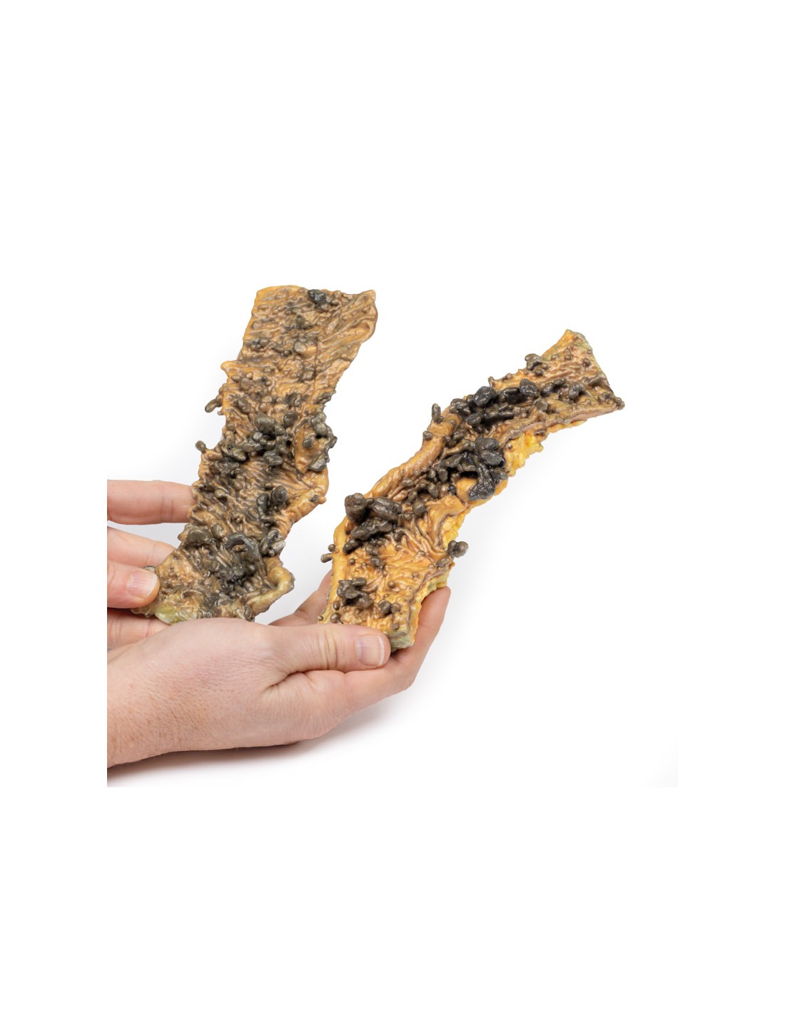
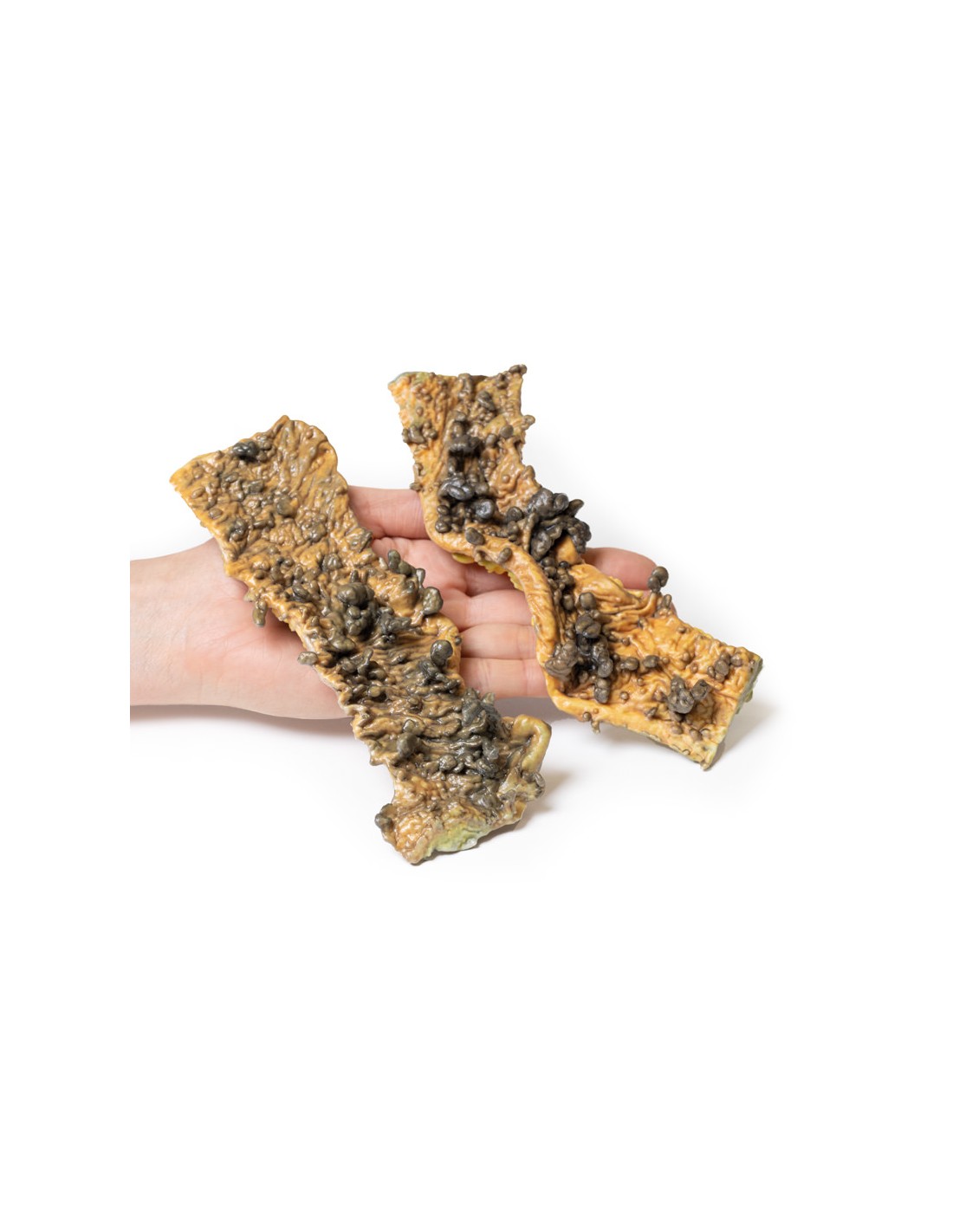
This dissection model highlighting multiple Polyposis is part of the exclusive Monash 3D anatomy series, a comprehensive series of human dissections reproduced with ultra-high resolution color 3D printing.
Clinical history.
There are no details of the clinical history.
Pathology
The specimens in this case consist of two segments of the sigmoid colon. The mucosa of the intestine is studded with numerous sessile and pedunculated partially pigmented polyps up to 1.5 cm in maximum diameter. There is no macroscopic evidence of malignant changes.
Note
Microscopically, polyps are most commonly tubular adenomas (>75% have a tubular structure; also called adenomatous polyps). Less frequently, they are villous adenomas (>75% have villous morphology) or tubulovilli adenomas (25-75% villous). They can have varying degrees of dysplasia. Histologic appearance is identical to sporadic colonic adenomas.
Patients with familial adenomatous polyposis (FAP; a form of hereditary colon cancer syndrome involving the APC gene located on chromosome 5q21) are offered prophylactic colectomy because it is almost certain that invasive adenocarcinoma will develop into one or more polyps, usually about 15 years after the onset of adenomatosis. This condition is transmitted as an autosomal dominant character.
What advantages does the Monash University anatomical dissection collection offer over plastic models or plastinated human specimens?
- Each body replica has been carefully created from selected patient X-ray data or human cadaver specimens selected by a highly trained team of anatomists at the Monash University Center for Human Anatomy Education to illustrate a range of clinically important areas of anatomy with a quality and fidelity that cannot be achieved with conventional anatomical models-this is real anatomy, not stylized anatomy.
- Each body replica has been rigorously checked by a team of highly trained anatomists at the Center for Human Anatomy Education, Monash University, to ensure the anatomical accuracy of the final product.
- The body replicas are not real human tissue and therefore not subject to any barriers of transportation, import, or use in educational facilities that do not hold an anatomy license. The Monash 3D Anatomy dissection series avoids these and other ethical issues that are raised when dealing with plastinated human remains.
No reviews
Tap to zoom