Your cart
There are no more items in your cart


Chronic gastric ulcers - Erler Zimmer 3D anatomy Series MP2076
erler zimmer
EZ-MP2076
€632.08
Tax included
Made in ultra-high resolution 3D printing in full color.
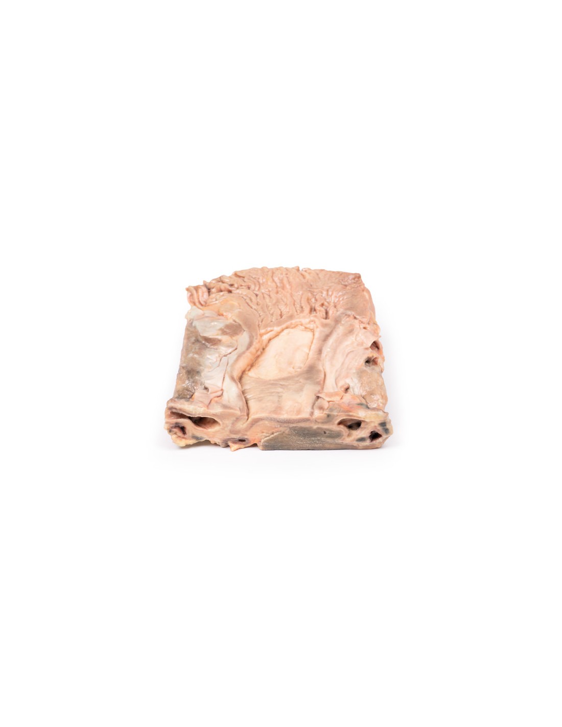
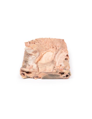
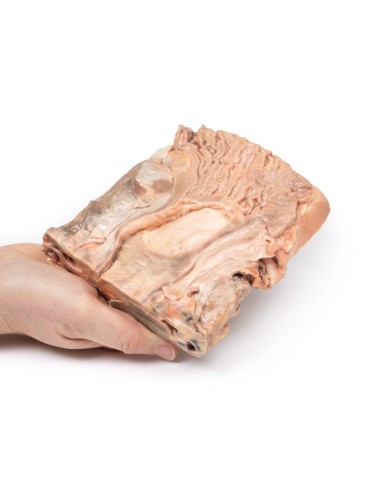
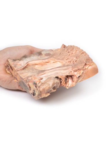
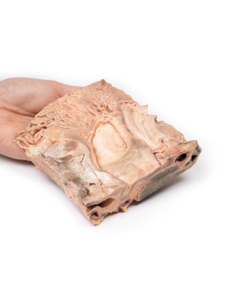
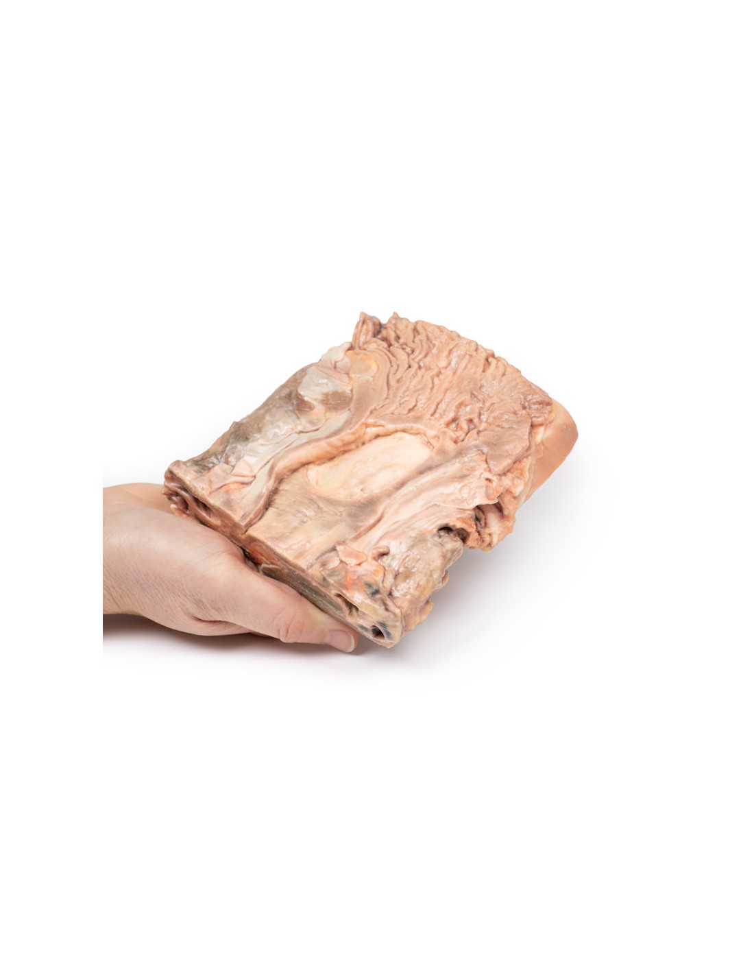
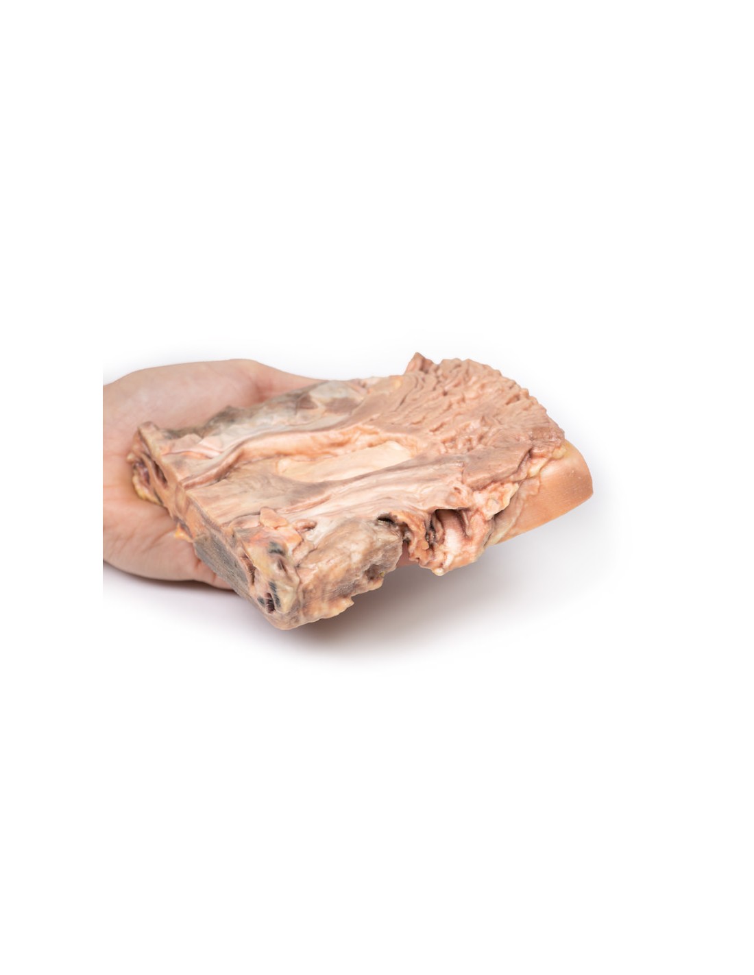
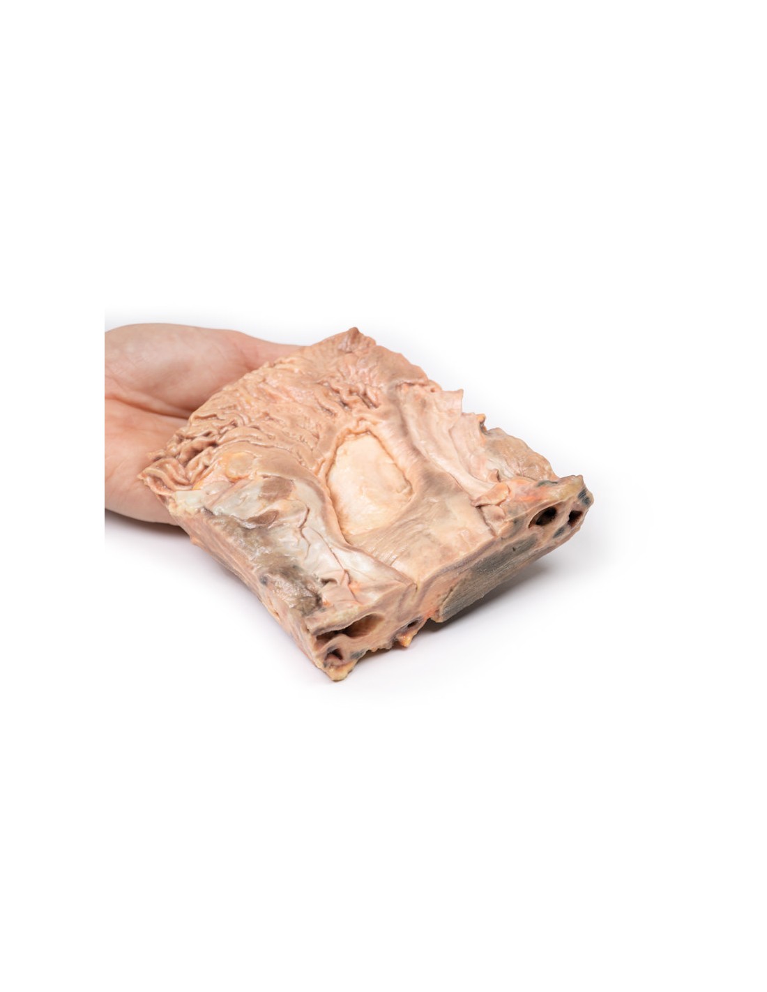
Chronic Gastric Ulcers - Erler Zimmer 3D anatomy Series MP2076
This dissection model highlighting a cholelithiasis is part of the exclusive Monash 3D anatomy series, a comprehensive series of human dissections reproduced with ultra-high resolution color 3D printing.
Clinical history
This elderly patient had a long history of "indigestion." He collapsed and died after a massive stroke.
Pathology
The specimen is a 2-cm slice of coronal tissue, incorporating a portion of the stomach diaphragm, liver, and pancreas. The specimen was opened to show a large ulcer at the upper end of the minor curvature near the gastroesophageal junction. Macroscopically, the loss of substance at the ulcer site is oval, 5-6 cm in diameter, and slightly raised edges. The base is clean and smooth with no evidence of hemorrhage. The gastric wall surrounding the ulcer is hardened, due to fibrosis involving the ulcer base and spreading below the surrounding mucosa. Being retractable, the fibrosis is able to "pull" the gastric mucosa toward the base of the ulcer, so that the folds of the gastric mucosa converge radially around the loss of substance (this feature is not found in ulcerated gastric malignancies).
Further information
Patients with a gastric ulcer may experience worsening pain while eating, often described as a burning or dull ache. Other symptoms include belching, vomiting, weight loss, or poor appetite. Complications may include bleeding, perforation, and stomach blockage. Common causes include Helicobacter pylori bacteria and nonsteroidal anti-inflammatory drugs (NSAIDs).
H. pylori was first identified as a cause of peptic ulcers by Barry Marshall and Robin Warren of the University of Western Australia in the late 20th century, a discovery for which they received the Nobel Prize in 2005. Other less common causes include tobacco smoking, stress due to serious illness, Behçet's disease, Zollinger-Ellison syndrome, Crohn's disease and liver cirrhosis[1]. Older people are more susceptible to the effects of NSAIDs causing ulcers[1].
Diagnosis is typically suspected because of presenting symptoms with confirmation by endoscopy or barium swallowing[1]. H. pylori can be diagnosed by testing antibodies in the blood, a urea breath test, testing stool for signs of the bacteria, or a stomach biopsy[1]. Other conditions that produce similar symptoms include stomach cancer, coronary artery disease, and inflammation of the stomach lining (gastritis) or inflammation of the gallbladder (cystitis)[1].
Treatment includes quitting smoking, stopping the use of NSAIDs, reducing or preferably stopping alcohol consumption, and taking medications to reduce stomach acidity[1]. Ulcers due to H. pylori are treated with a combination of drugs, such as amoxicillin, clarithromycin and a proton pump inhibitor (PPI). The drug used to decrease acid is usually a PPI or H2 blocker (histamine H2 receptor antagonists). Bleeding ulcers can be treated by endoscopy, with open surgery usually used only in cases where it is unsuccessful. Peptic ulcers occur in about 4% of the population[1].
Scientific references
1. Najm WI (2011). "Peptic ulcer disease. Primary Care. 38 (3): 383-94.
What advantages does the Monash University anatomical dissection collection offer over plastic models or plastinated human specimens?
- Each body replica has been carefully created from selected patient X-ray data or human cadaver specimens selected by a highly trained team of anatomists at the Monash University Center for Human Anatomy Education to illustrate a range of clinically important areas of anatomy with a quality and fidelity that cannot be achieved with conventional anatomical models-this is real anatomy, not stylized anatomy.
- Each body replica has been rigorously checked by a team of highly trained anatomists at the Center for Human Anatomy Education, Monash University, to ensure the anatomical accuracy of the final product.
- The body replicas are not real human tissue and therefore not subject to any barriers of transportation, import, or use in educational facilities that do not hold an anatomy license. The Monash 3D Anatomy dissection series avoids these and other ethical issues that are raised when dealing with plastinated human remains.
No reviews
Tap to zoom