
Your cart
There are no more items in your cart


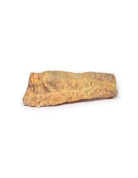
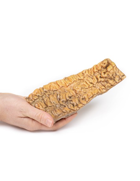
Ulcerative Colitis - Erler Zimmer 3D anatomy Series MP2074
erler zimmer
EZ-MP2074
€393.21
Tax included
Made in ultra-high resolution 3D printing in full color.
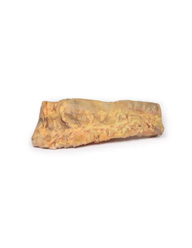
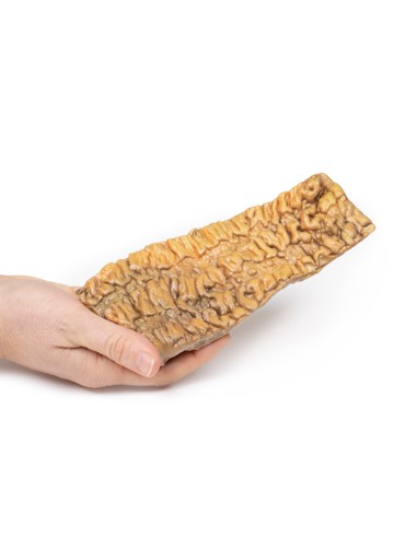
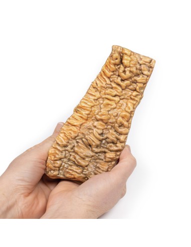
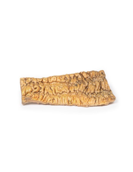
Ulcerative Colitis - Erler Zimmer 3D anatomy Series MP2074
This dissection model highlighting ulcerative colitis is part of the exclusive Monash 3D anatomy series, a comprehensive series of human dissections reproduced with ultra-high resolution color 3D printing.
Clinical History.
A 36-year-old woman was admitted to the hospital with a 3-week history of bloody diarrhea and lower abdominal pain. Further history revealed 4 similar episodes of diarrhea and abdominal pain in the past 7 years. Sigmoidoscopy showed erythematous, ulcerated and edematous rectal mucosa. She started steroid treatment but her symptoms did not improve. She underwent a total colectomy.
Pathology
The resected colon was cut open longitudinally to show the mucosal surface. There is extensive confluent ulceration separated by edematous islands of residual mucosa. The ulcerations have necrotic bases with prominent margins, some of which form "pseudo"-polypes. Histology of the intestinal mucosa showed acute inflammatory changes with crypt abscesses, focal necrosis, and ulcerations. This is an example of acute ulcerative colitis (UC).
Additional Information.
Ulcerative colitis is a chronic ulcerative-inflammatory disease that usually involves the rectum and may extend proximally in a continuous pattern to involve other parts of the colon. The inflammatory process is diffuse but is usually limited to the mucosa and superficial submucosa. The causative factor for UC is unknown. It most commonly begins between 15 and 25 years of age and is slightly more common in females.
Patients with CU may present with diarrhea, which may contain blood or mucus, fecal urgency, fecal frequency, tenesmus, abdominal pain with colic, as well as weight loss, anemia and fatigue. Inflammation of the colon can lead to toxic megacolon, colon perforation, or colon cancer. Extraintestinal manifestations include anterior uveitis, polyarthritis migrans, sacroiliitis, ankylosing spondylitis, erythema nodosum, pyoderma gangrenosum, and primary sclerosing cholangitis.
Treatments of CU include the use of anti-inflammatory drugs such as steroids, disease-modifying rheumatoid drugs and TNF (tumor necrosis factor) inhibitors. Colectomy essentially cures the intestinal symptoms of CU but extraintestinal manifestations of the disease may persist.
What advantages does the Monash University anatomical dissection collection offer over plastic models or plastinated human specimens?
- Each body replica has been carefully created from selected patient X-ray data or human cadaver specimens selected by a highly trained team of anatomists at the Monash University Center for Human Anatomy Education to illustrate a range of clinically important areas of anatomy with a quality and fidelity that cannot be achieved with conventional anatomical models-this is real anatomy, not stylized anatomy.
- Each body replica has been rigorously checked by a team of highly trained anatomists at the Center for Human Anatomy Education, Monash University, to ensure the anatomical accuracy of the final product.
- The body replicas are not real human tissue and therefore not subject to any barriers of transportation, import, or use in educational facilities that do not hold an anatomy license. The Monash 3D Anatomy dissection series avoids these and other ethical issues that are raised when dealing with plastinated human remains.
No reviews
Tap to zoom
