Your cart
There are no more items in your cart


Pedunculated adenoma of the colon - Erler Zimmer 3D anatomy Series MP2081
erler zimmer
EZ-MP2081
€252.30
Tax included
Made in ultra-high resolution 3D printing in full color.
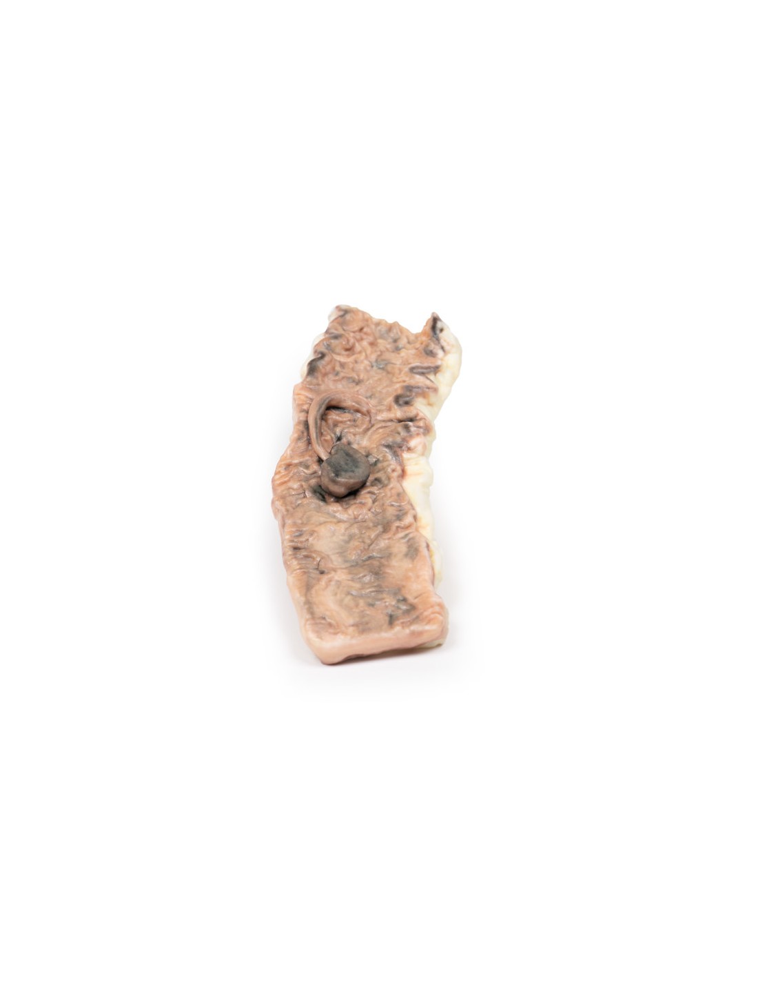
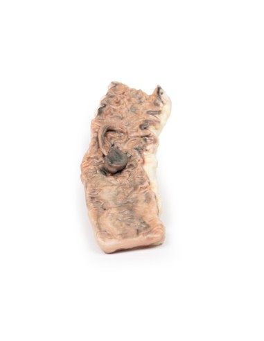
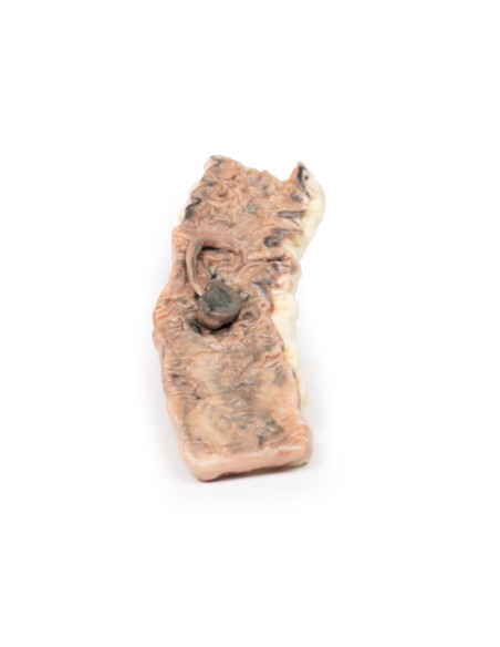
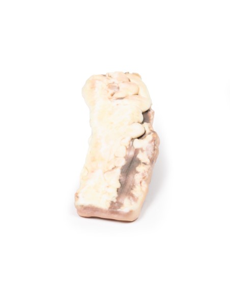
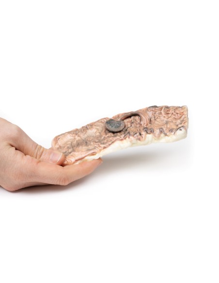
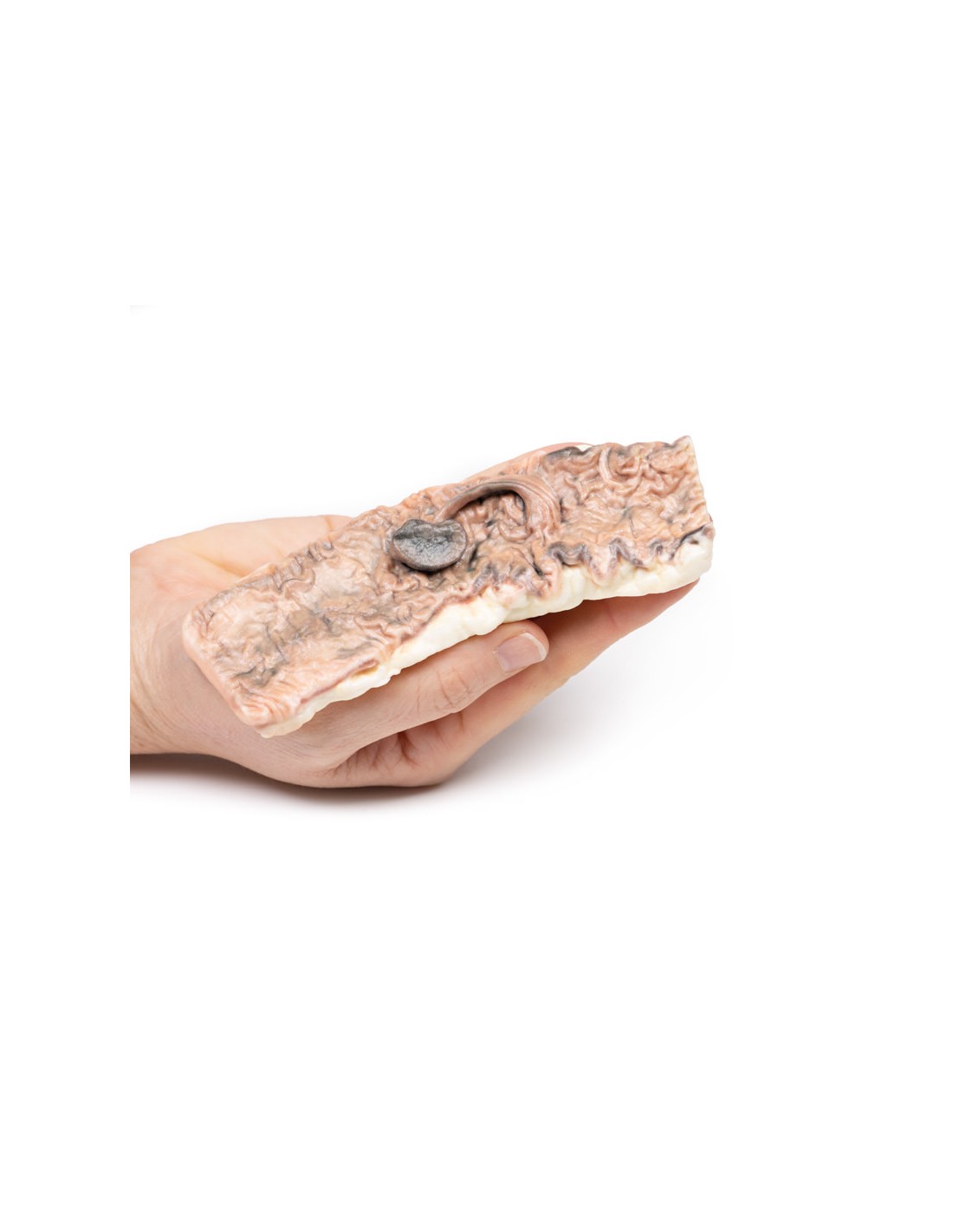
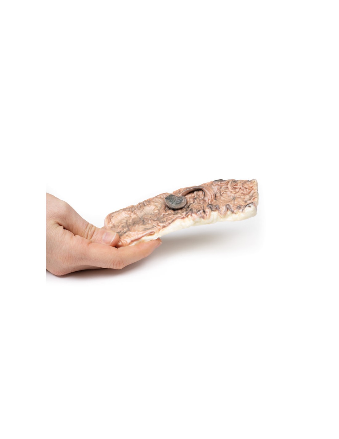
Pedunculated Adenoma of the Colon - Erler Zimmer 3D anatomy Series MP2081
This dissection model highlighting a Pedunculated Adenoma of the Colon? is part of the exclusive Monash 3D anatomy series, a comprehensive series of human dissections reproduced with ultra-high resolution color 3D printing.
Clinical History.
A 50-year-old man underwent colonoscopy after testing positive for fecal occult blood during a screening test. The colonoscopy revealed a pedunculated tumor in the descending colon, which was subsequently excised.
Pathology
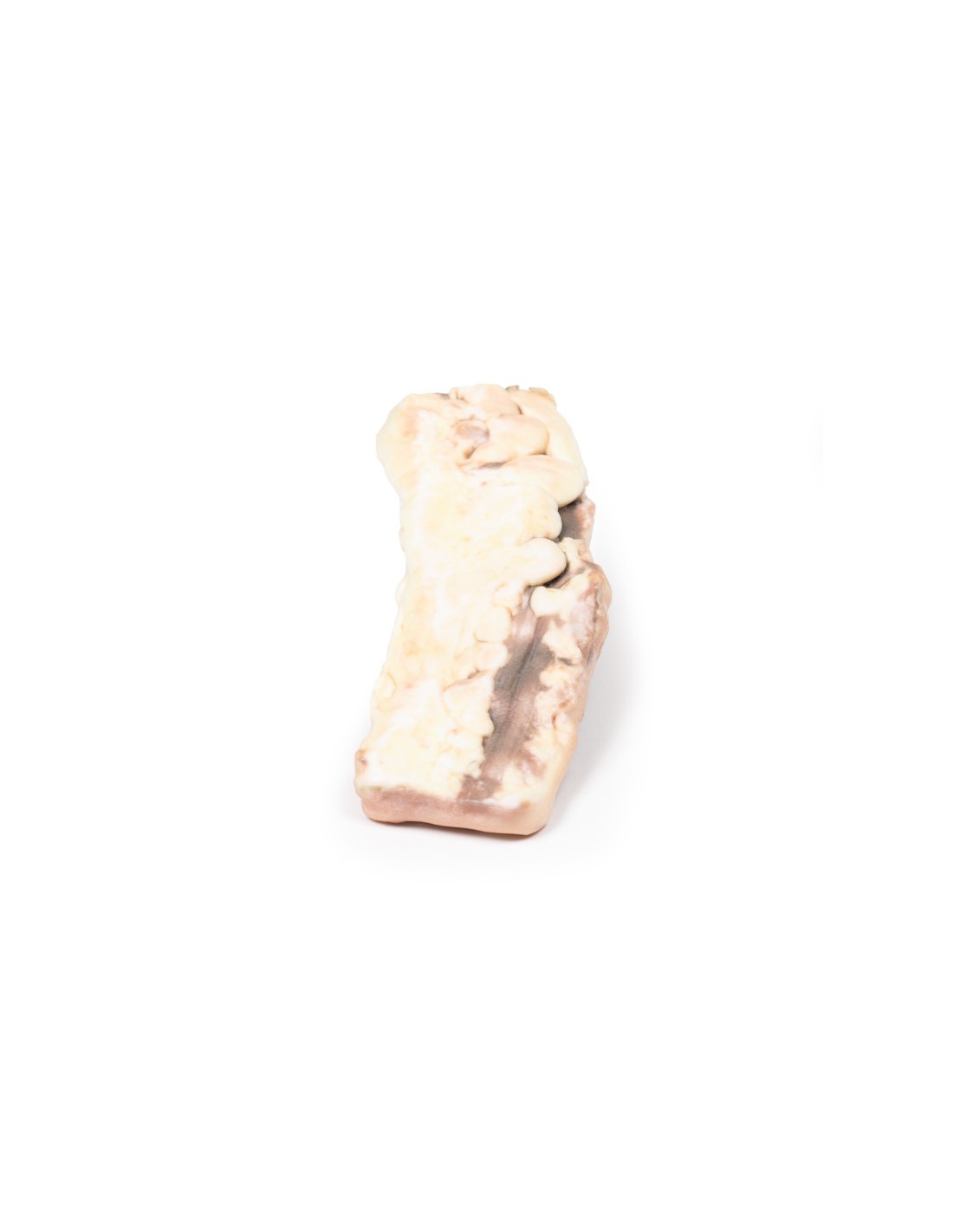
This specimen is the excised segment of the descending colon. There is a single visible dark lobulated mass arising from the mucosal surface. It is attached to a 4-cm-long stalk. Histologically, the mass comprises a connective tissue core covered by hyperplastic colon-like glandular epithelium with focal nuclear atypia. This is an example of tubular adenoma of the colon.
Additional Information.
Colorectal adenomas are intraepithelial neoplasms that characteristically exhibit epithelial dysplasia. They are benign but are precursors to adenocarcinoma. Not all adenomas evolve into adenocarcinoma. They produce polyps (sometimes pedunculated) or sessile or variable-sized lesions. They occur predominantly in males and are more common in Western countries due to diet and lifestyle. They are present in about 30% of people over 60 years of age in the West. There is an increased risk in patients with a positive family history of colorectal adenocarcinoma. Regular surveillance colonoscopy in risk groups with polyp removal reduces the incidence of adenocarcinoma. There are three classifications of colonic adenomas based on their architecture: tubular (>75% have tubular morphology), tubulovillus (25-75% villous morphology), and villous (>75% have villous morphology). Histologically, they may present epithelial dysplasia characterized by nuclear hyperchromasia, elongation, and stratification. Tubular adenomas tend to be small pedunculated polygons composed of rounded or tubular glands. Pedunculated adenomas have a thin fibromuscular stalk with blood vessels derived ??from the submucosa. The stalk is usually nonneoplastic epithelium. Adenoma size is the greatest predictor of progression to adenocarcinoma. Progression is rare in adenomas <1 cm in diameter. However, up to 40% of lesions greater than 4 cm in diameter evolve into adenocarcinoma. pedunculated polygons composed of round or tubular glands. Pedunculated adenomas have a thin fibromuscular stalk with blood vessels derived ??from the submucosa. The stalk is usually nonneoplastic epithelium. Adenoma size is the greatest predictor of progression to adenocarcinoma. Progression is rare in adenomas <1 cm in diameter. However, up to 40% of lesions greater than 4 cm in diameter evolve into adenocarcinoma. pedunculated polygons composed of round or tubular glands. Pedunculated adenomas have a thin fibromuscular stalk with blood vessels derived ??from the submucosa. The stalk is usually nonneoplastic epithelium. Adenoma size is the greatest predictor of progression to adenocarcinoma. Progression is rare in adenomas <1 cm in diameter. However, up to 40% of lesions greater than 4 cm in diameter evolve into adenocarcinoma.
Most adenomas are asymptomatic and slow-growing. Large polyps may present with symptoms of occult bleeding anemia. Villi adenomas occasionally secrete large amounts of protein ??mucoid and/or potassium-rich fluids, possibly leading to hypokalemia.
.
What advantages does the Monash University anatomical dissection collection offer over plastic models or plastinated human specimens?
- Each body replica has been carefully created from selected patient X-ray data or human cadaver specimens selected by a highly trained team of anatomists at the Monash University Center for Human Anatomy Education to illustrate a range of clinically important areas of anatomy with a quality and fidelity that cannot be achieved with conventional anatomical models-this is real anatomy, not stylized anatomy.
- Each body replica has been rigorously checked by a team of highly trained anatomists at the Center for Human Anatomy Education, Monash University, to ensure the anatomical accuracy of the final product.
- The body replicas are not real human tissue and therefore not subject to any barriers of transportation, import, or use in educational facilities that do not hold an anatomy license. The Monash 3D Anatomy dissection series avoids these and other ethical issues that are raised when dealing with plastinated human remains.
No reviews
Tap to zoom