Your cart
There are no more items in your cart


Chronic hydrocele - Erler Zimmer 3D anatomy Series MP2110
erler zimmer
EZ-MP2110
€280.48
Tax included
Made in ultra-high resolution 3D printing in full color.
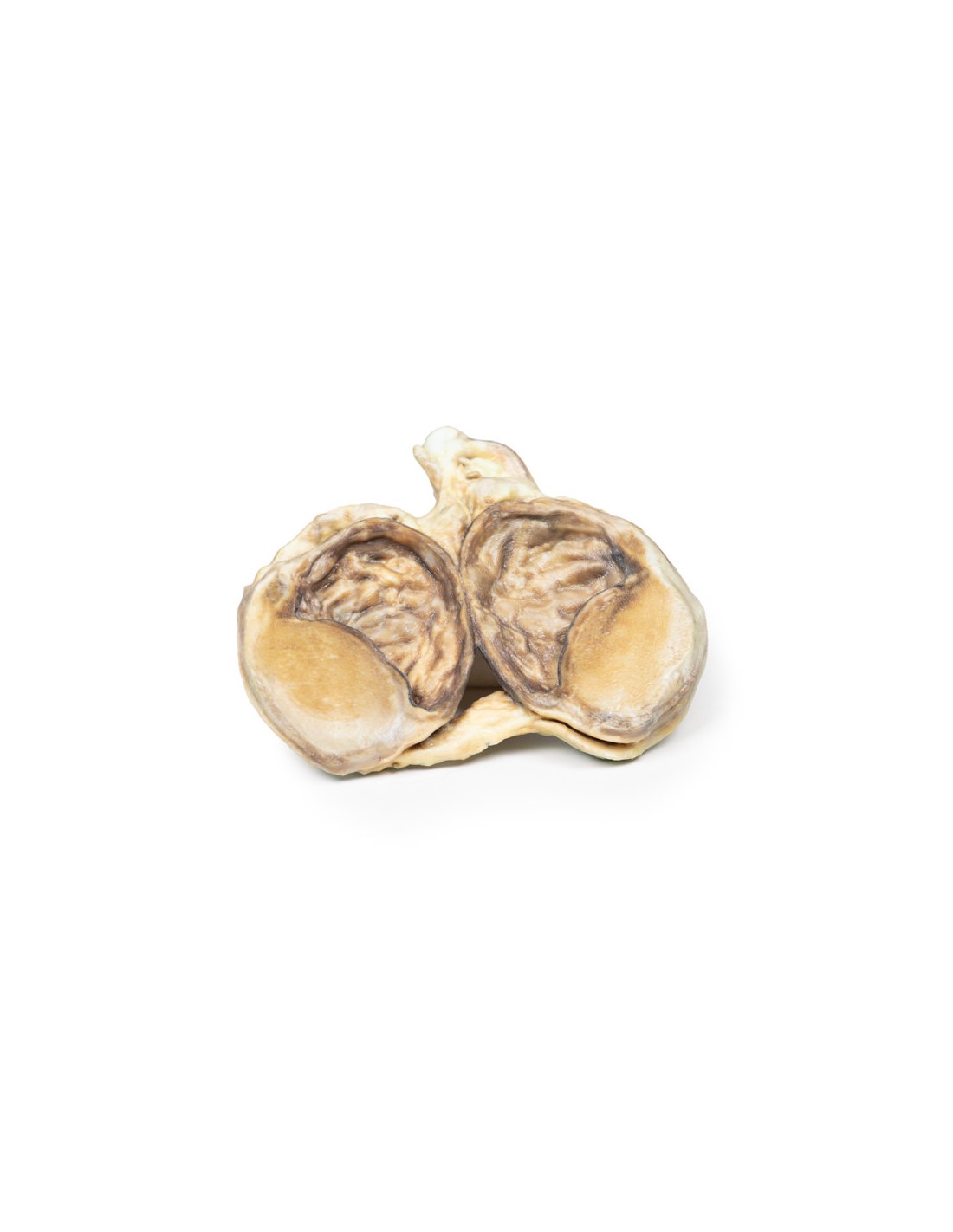
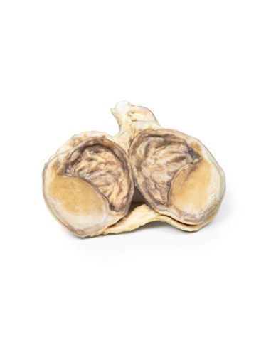
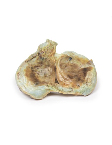
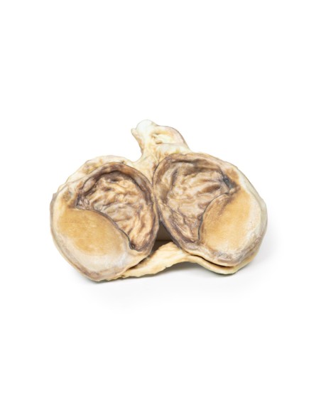
Chronic Hydrocele - Erler Zimmer 3D anatomy Series MP2110
This dissection model highlighting a chronic Hydrocele is part of the exclusive Monash 3D anatomy series, a comprehensive series of human dissections reproduced with ultra-high resolution color 3D printing.
Clinical history
Of an 80-year-old man presented with hematemesis. He has a known history of alcoholic liver cirrhosis with esophageal varices. On examination, it is noted that he has multiple spider nevi, large volume abdominal ascites, and scrotal swelling. Transillumination of the swelling transmitted red light. He had another large-volume hematemesis and died shortly after admission.
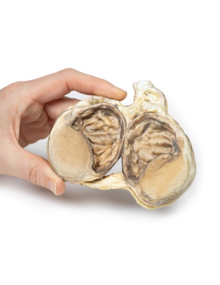
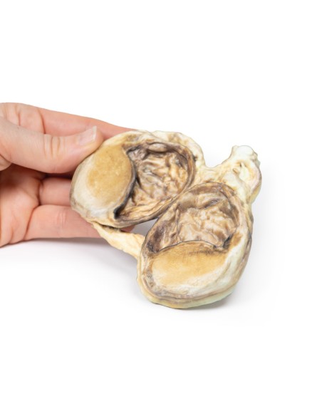
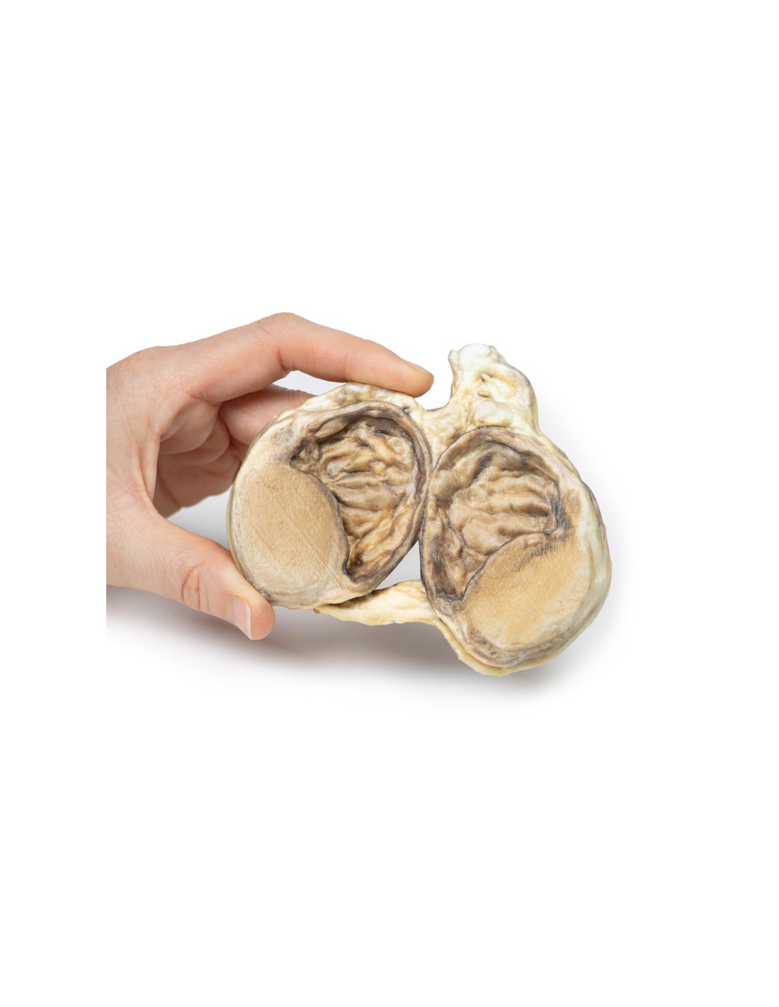
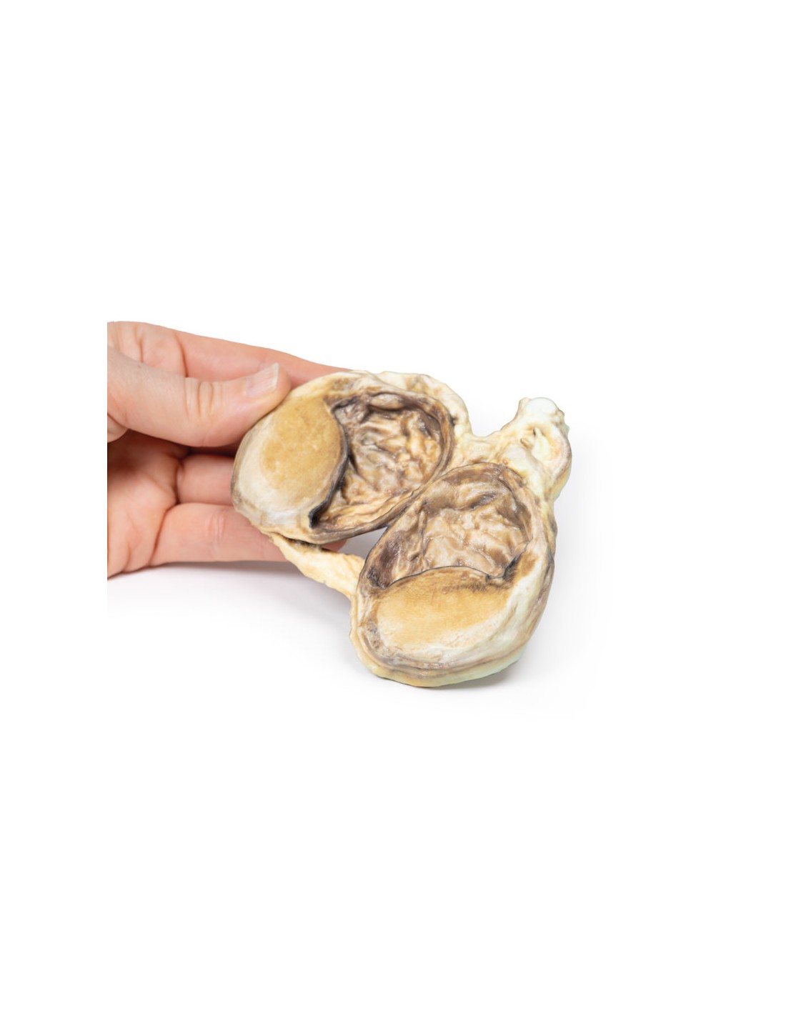
Pathology
The specimen consists of testis, tunica vaginalis, and distal end of the spermatic cord. The testis and surrounding layers were cut in two to visualize the cut surface. The tunica vaginalis is thickened and the closed cavity is dilated. The testis is normal. This is an example of chronic secondary communicated hydrocele.
Additional Information.
A hydrocele is an accumulation of serous fluid between the parietal and visceral layers of the tunica vaginalis around the testes. Hydrocele can be described as communicating with the peritoneal cavity or not communicating with the peritoneal cavity.
Communicating hydrocele develops due to the failure of the processus vaginalis to close after the tests descend into the scrotum. These can occur after birth as congenital hydrocele or can occur later in life due to increased intra-abdominal pressure, such as heart failure in this case. Noncommunicating hydroceles are caused by imbalances in fluid secretion and reabsorption (e.g., orchitis, epididymitis), testicular tumor, physical trauma (e.g., hernia, testicular torsion) or defective lymphatic drainage (e.g., filariasis, elephantiasis).
Patients present with a scrotal mass. The mass may be uni- or bilateral. Communicating hydrocele may be reducible and increase in size with increasing intra-abdominal pressure. Noncommunicating are generally nonreducible swellings. The swelling is usually not painful unless there is an underlying infection or torsion causing the hydrocele. Larger hydroceles can be bulky and cause erosion and infection of the skin on the scrotum.
Diagnosis can be made on objective examination. Serous fluid allows shining light to pass through the scrotum when it is examined: this is called transillumination. Ultrasound can be used to consolidate the diagnosis and rule out other testicular pathologies. Serum markers of testicular cancer, such as alpha fetoprotein and B-HCG, can be taken to rule out testicular cancer.
Many congenital hydroceles resolve spontaneously before the age of 2 years. If communicating hydroceles persist beyond 2 years, they are repaired surgically because of the risk of developing incarcerated hernias. Surgical repair of communicating hydrocele in older patients may be offered if they are symptomatic. Treatment of the underlying etiology of reactive hydrocele can cause its resolution.
What advantages does the Monash University anatomical dissection collection offer over plastic models or plastinated human specimens?
- Each body replica has been carefully created from selected patient radiographic data or human cadaver specimens selected by a highly trained team of anatomists at the Monash University Center for Human Anatomy Education to illustrate a range of clinically important areas of anatomy with a quality and fidelity that cannot be achieved with conventional anatomical models-this is real anatomy, not stylized anatomy.
- Each body replica has been rigorously checked by a team of highly trained anatomists at the Center for Human Anatomy Education, Monash University, to ensure the anatomical accuracy of the final product.
- The body replicas are not real human tissue and therefore not subject to any barriers of transportation, import, or use in educational facilities that do not hold an anatomy license. The Monash 3D Anatomy dissection series avoids these and other ethical issues that are raised when dealing with plastinated human remains.
No reviews
Tap to zoom