Your cart
There are no more items in your cart


Nodular hyperplasia of the prostate - Erler Zimmer 3D anatomy Series MP2108
erler zimmer
EZ-MP2108
€203.98
Tax included
Made in ultra-high resolution 3D printing in full color.
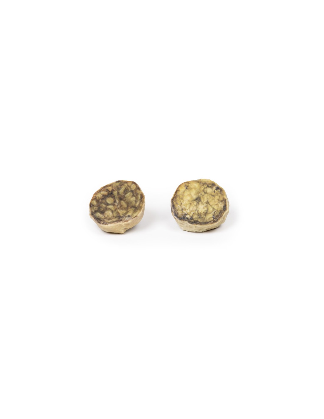
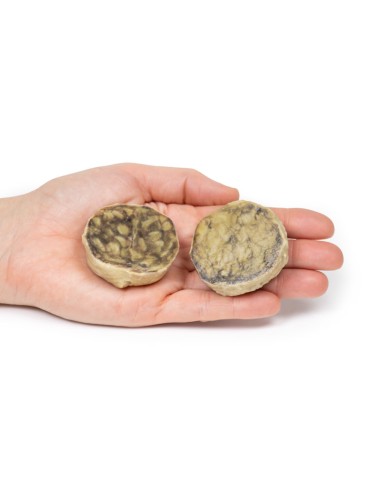
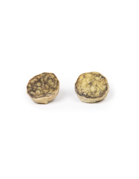
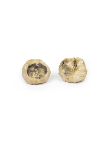
Nodular Hyperplasia of the Prostate - Erler Zimmer 3D anatomy Series MP2108
This dissection model highlighting Nodular Hyperplasia of the Prostate is part of the exclusive Monash 3D anatomy series, a comprehensive series of human dissections reproduced with ultra-high resolution color 3D printing.
Clinical history
A 63-year-old man presented to the emergency department started with acute abdominal pain. He was unable to urinate for 5 days. Further questioning revealed a 2-year history of urinary frequency, double urination, urinary hesitation, nocturia, and poor urine flow. Abdominal examination showed a tender distended bladder and an enlarged palpable prostate on digital rectal examination. A bedside bladder scan demonstrated a volume of > 1 L in the bladder. Blood tests show severe acute kidney injury. He is diagnosed with acute renal failure due to acute urinary retention. There have been multiple unsuccessful attempts to catheterize the patient through the urethra and in an overdose manner. A total prostatectomy was performed and he recovered well.
Pathology
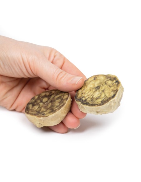
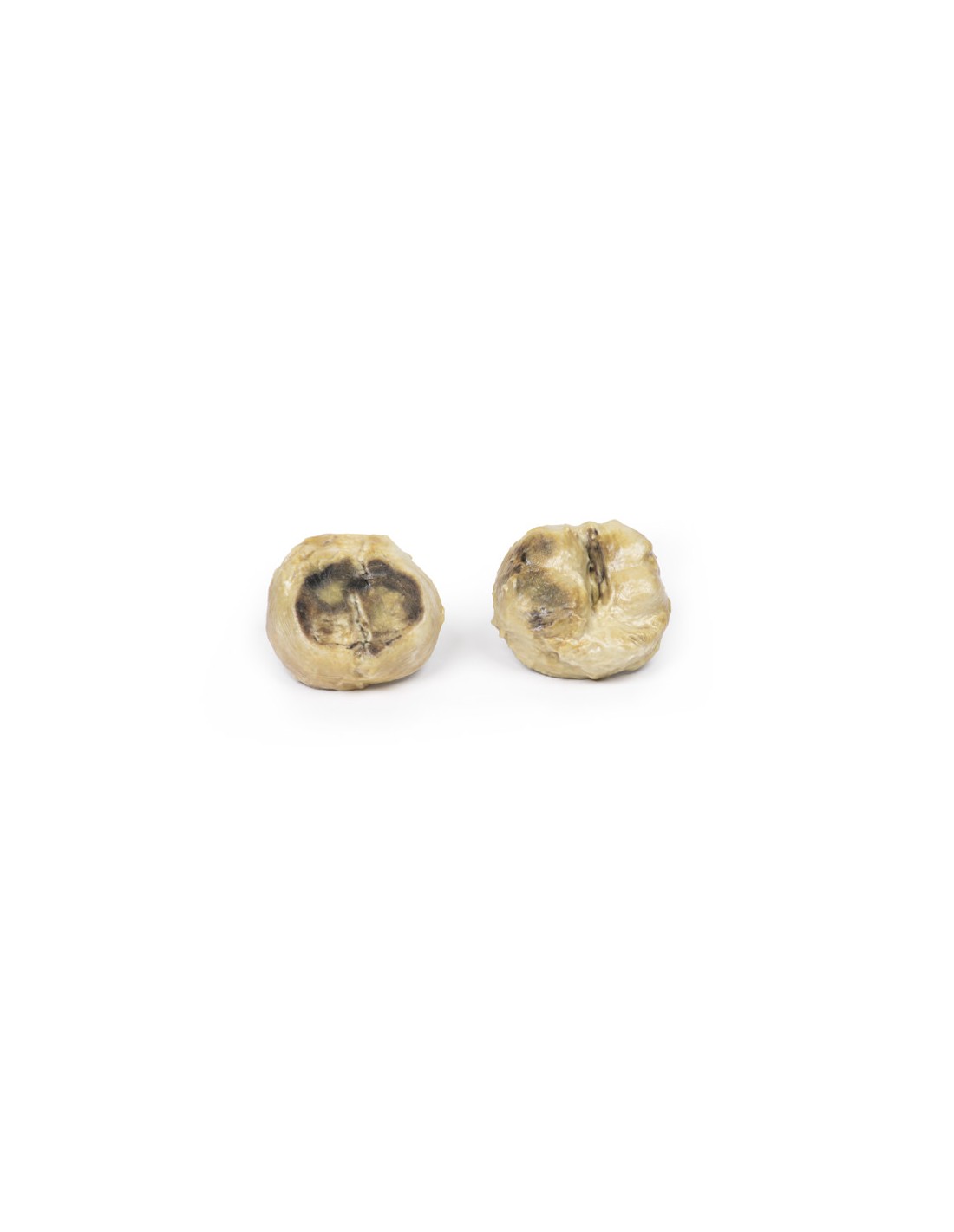
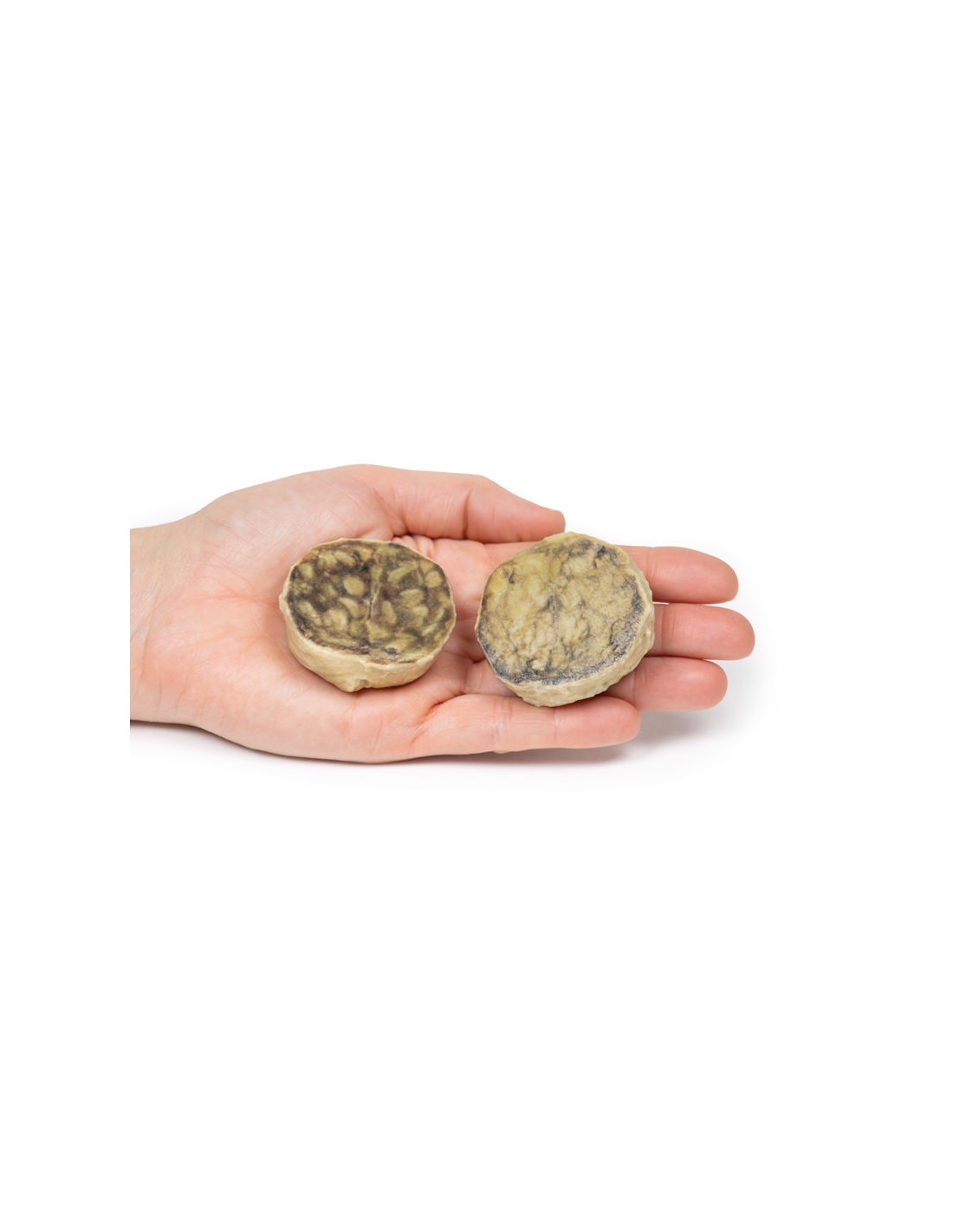
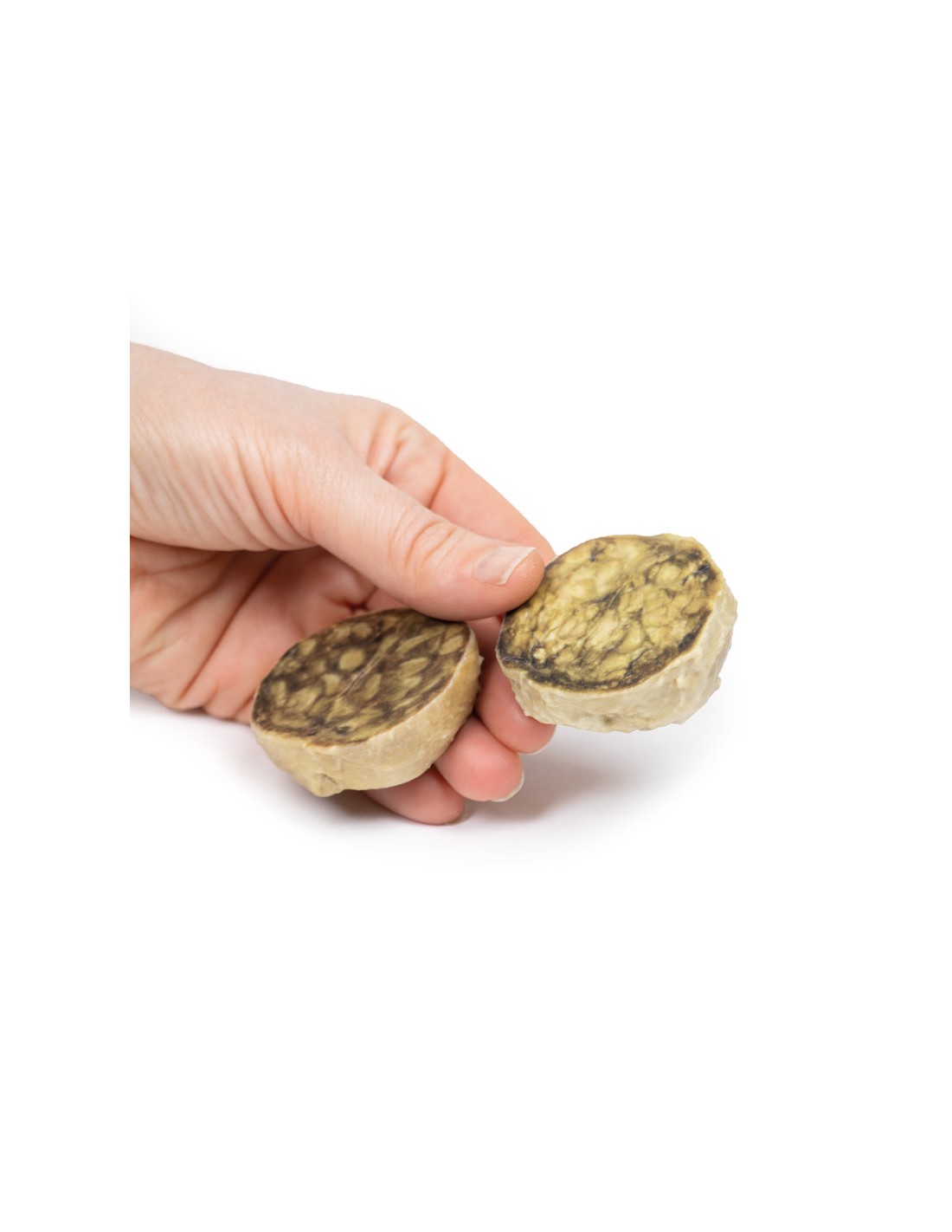
The specimen is an enlarged prostate gland sliced transversely to visualize the outer surfaces and cut, On the cut surface are numerous nodules ranging in size from 2-10 mm in diameter. This is an example of benign nodular hyperplasia (BPH) of the prostate gland.
Notes on Nodular Hyperplasia of the Prostate.
Benign prostatic hyperplasia (BPH) or nodular hyperplasia of the prostate is a common disease in older men. Benign prostatic hyperplasia is caused by nodular hyperplasia of prostatic stromal and glandular epithelial cells mainly in the periurethral prostate. Hyperplasia results from the accumulation of senescent cells due to reduced cell death and cell proliferation driven by androgens, mainly dihydrotestosterone. Disproportionate enlargement of the median lobe is a common feature of nodular hyperplasia of the prostate. The protruding median lobe may occlude the internal urethral orifice upon bladder contraction.
The prevalence of BPH increases significantly with age. BPH is present in 20% of males at age 40 years, in 70% of males aged 60 years, and in nearly 90% of males aged 80 years and older. There is an increased risk of BPH in men with a positive family history of BPH, obese males, and exposure to exogenous androgenic-anabolic steroids.
The clinical presentation of BPH results from obstructive urinary symptoms. Patients complain of urinary frequency, nocturia, urinary hesitation, double urination, poor urinary flow, and overflow drip. Acute urinary retention may result from complete urinary tract obstruction as in the case discussed above. Post-minctional residual urine results from prostate obstruction leading to an increased risk of urinary tract infections.
Diagnosis can be made on clinical history and physical examination of the prostate with a digital rectal examination. Prostate-specific antigen can be used to screen for prostate cancer. Ultrasound or CT scan can be used to assess prostate volume. BPH can be medically treated with alpha-blockers to relax prostate smooth muscle tone or 5-alpha-reductase inhibitors, which inhibit dihydrotesterone synthesis. The main surgical treatment for severe cases of BPH is transurethral resection of the prostate (TURP). Total prostatectomy is no longer used because of the risk of disabling complications.
What advantages does the Monash University anatomical dissection collection offer over plastic models or plastinated human specimens?
- Each body replica has been carefully created from selected patient X-ray data or human cadaver specimens selected by a highly trained team of anatomists at the Monash University Center for Human Anatomy Education to illustrate a range of clinically important areas of anatomy with a quality and fidelity that cannot be achieved with conventional anatomical models-this is real anatomy, not stylized anatomy.
- Each body replica has been rigorously checked by a team of highly trained anatomists at the Center for Human Anatomy Education, Monash University, to ensure the anatomical accuracy of the final product.
- The body replicas are not real human tissue and therefore not subject to any barriers of transportation, import, or use in educational facilities that do not hold an anatomy license. The Monash 3D Anatomy dissection series avoids these and other ethical issues that are raised when dealing with plastinated human remains.
No reviews
Tap to zoom