Your cart
There are no more items in your cart


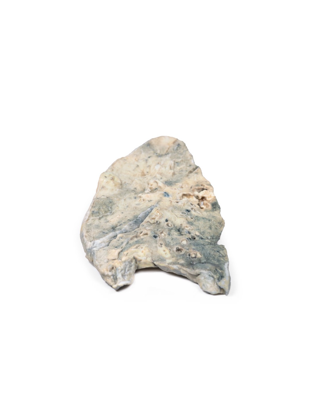
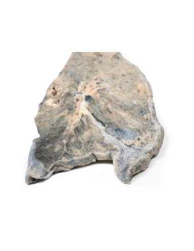
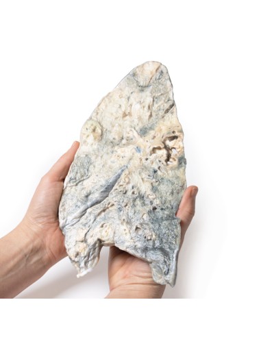
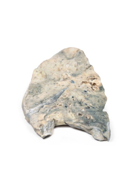
Lung: staphylococcus aureus abscess - Erler Zimmer 3D anatomy Series MP2064
erler zimmer
EZ-MP2064
€782.39
Tax included
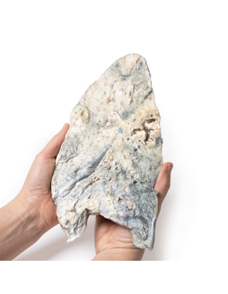
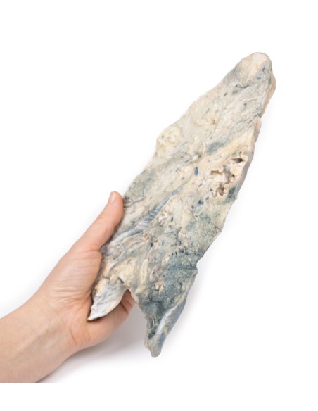
Made in ultra-high resolution 3D printing in full color.
Lung: staphylococcus aureus abscess - Erler Zimmer 3D anatomy Series MP2064
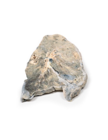
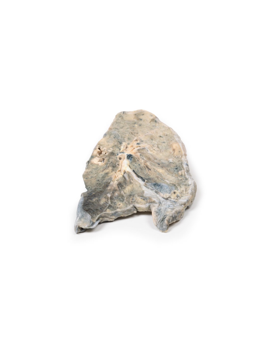
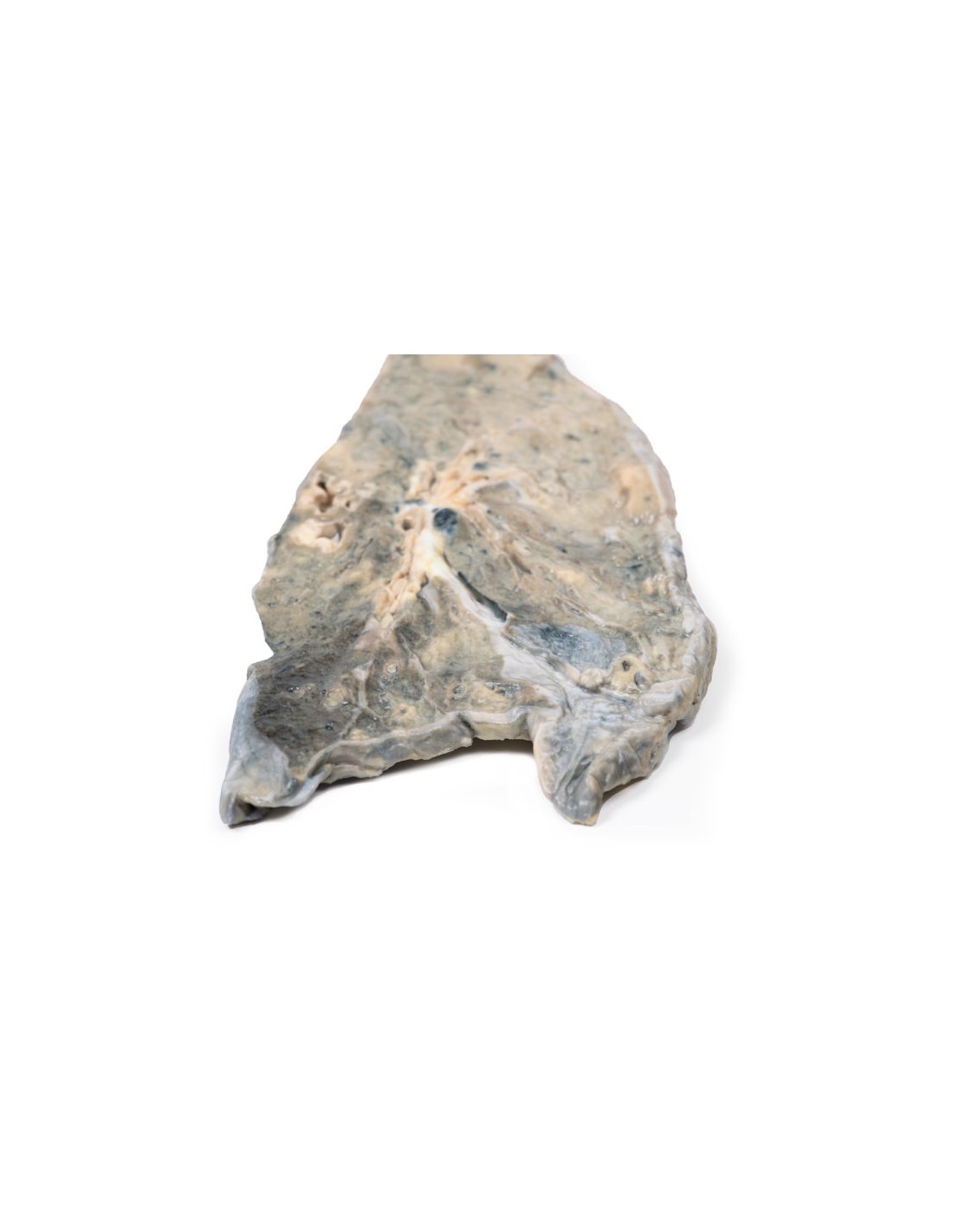
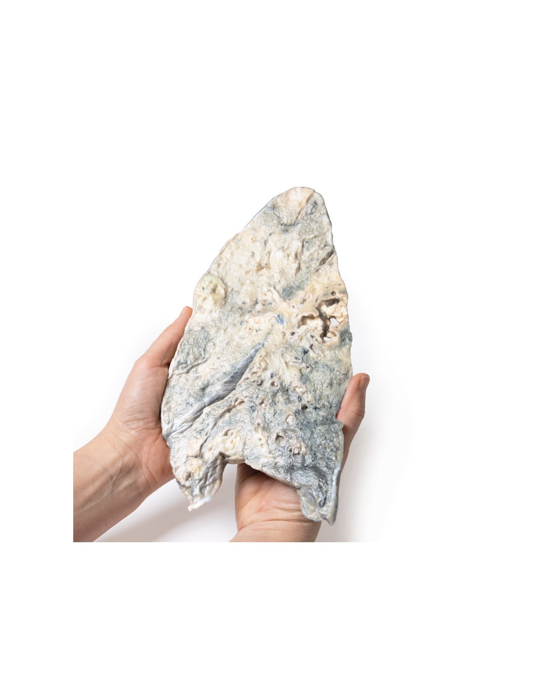
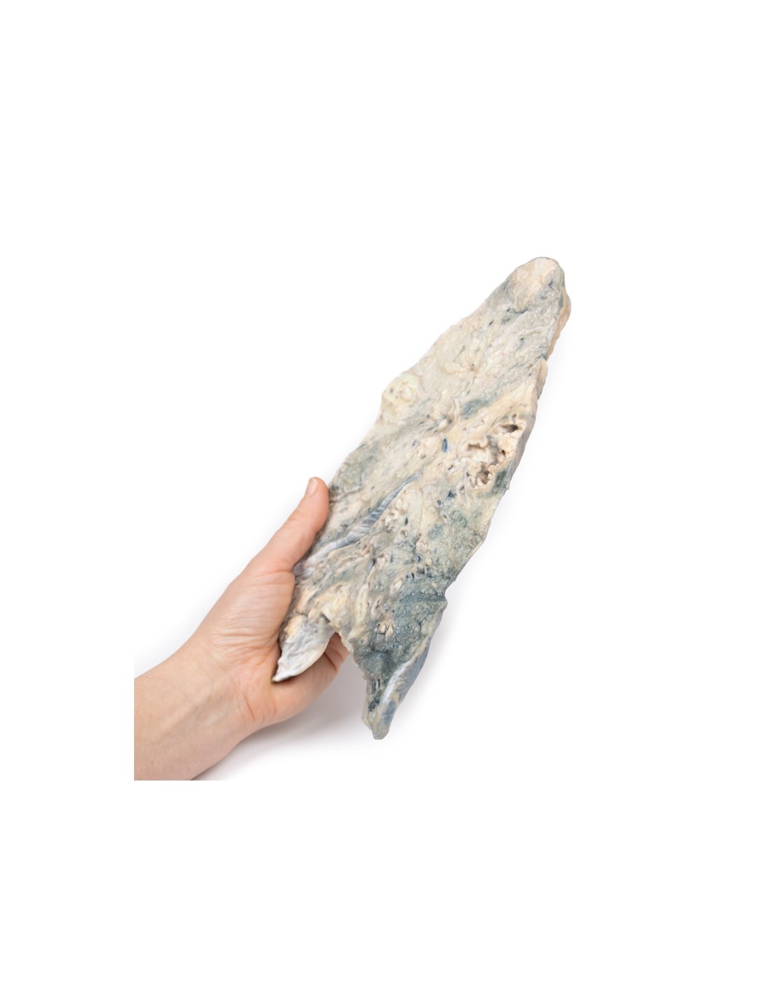
This dissection model highlighting a Lung with staphylococcus aureus abscess is part of the exclusive Monash 3D anatomy series, a comprehensive series of human dissections reproduced with ultra-high resolution color 3D printing.
Clinical history
A 55-year-old woman presents with severe dyspnea, productive cough and oral candidiasis. She is immunodepressed with a history of rheumatoid arthritis being treated with steroids and cyclophosphamide. Sputum cultures developed staphylococcus aureus. Appropriate therapy was initiated but she died shortly after admission.
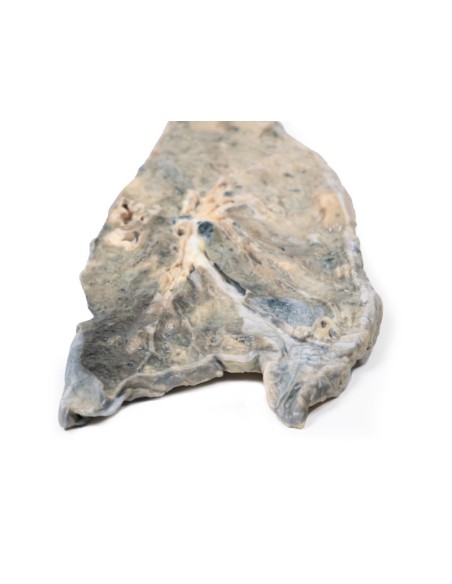
Pathology
The right lung was divided in two. Multiple irregular abscess cavities are visible. The largest of these, at the apex of the lower lobe, measures 4 x 3 cm in diameter. At the apex of the upper lobe, there is another, less obvious irregular abscess cavity measuring about 3 x 2 cm in diameter surrounded by an area of consolidation. A few small abscesses are also observed. Patchy consolidation is present in the middle lobe. Several bronchi contain and are obstructed with pus plugs. Cultures taken from the specimen are grown Staphylococcus. halo. This is an example of multiple staphylococcal lung abscesses in an immunosuppressed patient.
Additional Information.
Staphylococcus aureus is a gram-positive cocci. It is part of the microbiota of the human body usually found on the skin or upper respiratory tract. It is usually commensal but can cause opportunistic infections such as common or less common skin infections, pneumonia, and endocarditis. It can cause both community and nosocomial pneumonia. Hospital-acquired Staphylococcal pneumonia is most commonly associated with intubation and prolonged hospitalizations. The prevalence of hospital-acquired pneumonia caused by methicillin-resistant Staph Aureus (MRSA) is increasing.
It is an important cause of secondary bacterial pneumonia in patients following a viral respiratory infection, such as after a flu infection. Intravenous drug users have an increased risk of developing "metastatic" Staphylococcus. aureus pneumonia and endocarditis, following staphylococcal bacteremia caused by the use of dirty needles. Staph. aureus pneumonia is severe and associated with an increased rate of complications, such as cavity abscess formation and empyema.
Staph Aureus pneumonia should be suspected in any of the above high-risk groups, as well as in patients with rapidly deteriorating pneumonia, hemoptysis, early multilobar pneumonia on X-ray, pulmonary cavitation, or disseminated intravascular coagulation. First-line treatment for Staph. aureus pneumonia is a penicillin antibiotic, such as flucloxacillin. Staphylococcal resistance to penicillin is very common with the production of penicillinases, e.g., MRSA. MRSA is treated with glycopeptide antibiotics, such as vancomycin, or oxazolidinone antibiotics, such as linezolid.
What advantages does the Monash University anatomical dissection collection offer over plastic models or plastinated human specimens?
- Each body replica has been carefully created from selected patient X-ray data or human cadaver specimens selected by a highly trained team of anatomists at the Monash University Center for Human Anatomy Education to illustrate a range of clinically important areas of anatomy with a quality and fidelity that cannot be achieved with conventional anatomical models-this is real anatomy, not stylized anatomy.
- Each body replica has been rigorously checked by a team of highly trained anatomists at the Center for Human Anatomy Education, Monash University, to ensure the anatomical accuracy of the final product.
- The body replicas are not real human tissue and therefore not subject to any barriers of transportation, import, or use in educational facilities that do not hold an anatomy license. The Monash 3D Anatomy dissection series avoids these and other ethical issues that are raised when dealing with plastinated human remains.
No reviews
Tap to zoom