Your cart
There are no more items in your cart


Tracheoesophageal fistula and esophageal atresia - Erler Zimmer 3D anatomy Series MP2059
erler zimmer
EZ-MP2059
€232.17
Tax included
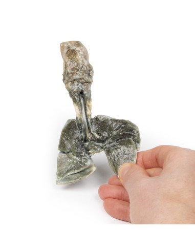
Made in ultra-high resolution 3D printing in full color.
Tracheoesophageal Fistula and Esophageal Atresia - Erler Zimmer 3D anatomy Series MP2059
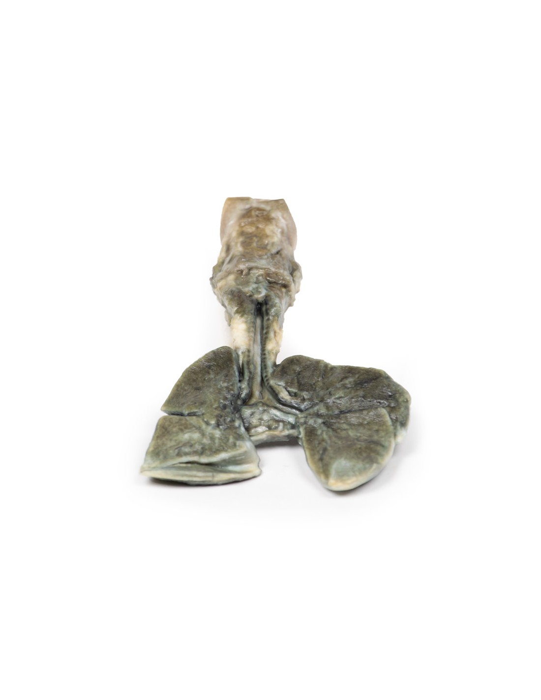
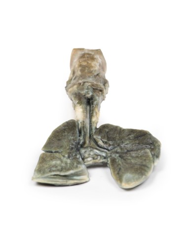
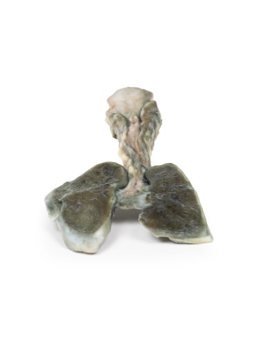
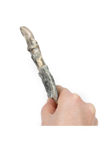
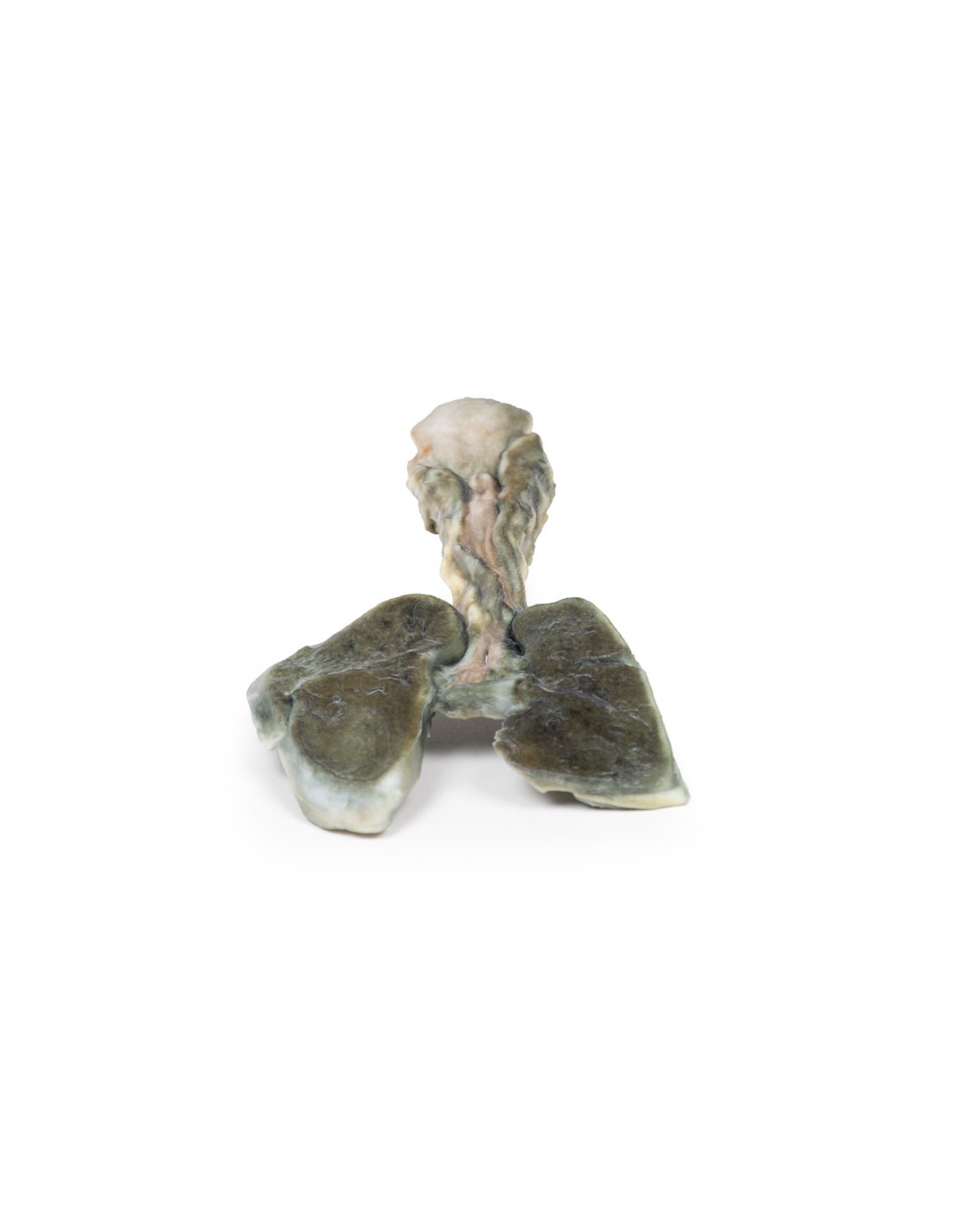
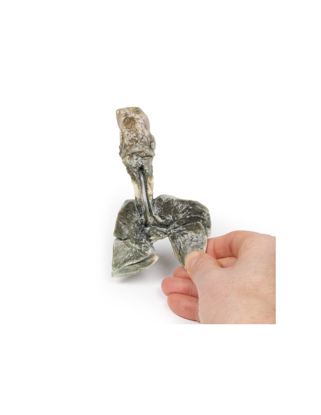
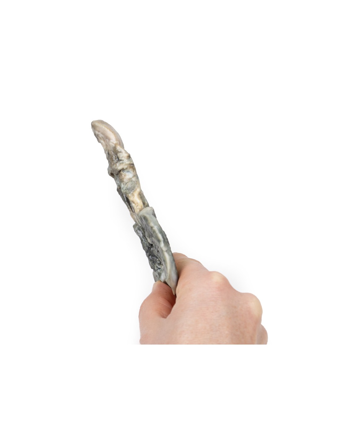
This dissection model highlighting a Tracheoesophageal Fistula and Esophageal Atresia is part of the exclusive Monash 3D anatomy series, a comprehensive series of human dissections reproduced with ultra-high resolution color 3D printing.
Clinical History.
A 32-year-old G3P0 woman (gravida 3, para 0' - that is, she had two pregnancies, with neither embryo surviving to a gestational age of 24 weeks) presented in preterm labor at 25 weeks' gestation. The physician had noted an increase in fundal height to 30 cm a week earlier, but the mother had refused prenatal testing or ultrasound and was lost to follow-up.She gave birth to a live-born male child. Examination of the baby revealed polydactyly, imperforate anus, excessive salivation, and a loud pan-systolic murmur. A single umbilical artery was detected in the umbilical cord. The child had difficulty feeding with increasing respiratory distress. The child died 2 days later from aspiration pneumonia.
Pathology/Details of the specimen
The specimen includes the tongue, larynx, trachea, bronchi, both lungs, and esophagus of the fetus. The trachea and bronchi were divided at the midline. A fistula is present just above the bifurcation at a communicating fistula connecting the distal esophagus to the trachea (arrow). This is an example of a type C tracheoesophageal fistula (esophageal atresia with distal tracheoesophageal fistula). It is difficult to discern whether the esophagus ends as a blind pouch in the lower part of the specimen.
Further information
Tracheoesophageal fistula (TEF) is a common congenital anomaly that occurs in about 1 in 4000 live births. FET usually occurs with esophageal atresia (sometimes abbreviated to EA, reflecting the U.S. spelling of "esophagus"). FETs are classified according to their anatomical configuration. Type C is the most common configuration; as described above, in which esophageal atresia with distal tracheoesophageal fistula constitutes 86% of cases. FET occurs without esophageal atresia in only 4% of cases, type E.
TEF and esophageal atresia are caused by a defective lateral setting of the anterior bowel in the esophagus and trachea. It is believed that a defect in epithelial-mesenchymal interactions causes a branch of the pulmonary bud to fail to branch, which becomes the fistula tract. It is associated with VACTERL (vertebral defects, anal atresia, heart defects, FET, renal abnormalities, and limb abnormalities) or CHARGE syndrome (Coloboma, heart defects, Atresia choanae, growth retardation, genital abnormalities, and ear abnormalities).
Esophageal atresia may be observed on prenatal ultrasonography as polydramnios, absent/collapsed stomach, and dilatation of the proximal esophageal pouch. EA with FET may be more difficult to see on ultrasound because the fistula allows fluid to flow into the stomach. Polydramnios occurs in one third of cases of EA with distal FET. Postnatal symptoms vary depending on the configuration of the fistula. These include excessive salivation, respiratory distress, difficulty feeding and choking. Reflux of gastric contents can lead to aspiration of pneumonia as in this case.
Diagnosis can be made by not passing a nasogastric tube into the stomach along with X-ray imaging. Fluoroscopy with contrast can be used for more indeterminate cases. For milder cases, diagnosis can be made later with endoscopic investigation. Treatment involves surgical correction of defects. The prognosis is generally good. However, cases with associated chromosomal, premature, and cardiac defects are at increased risk of death.
What advantages does the Monash University anatomical dissection collection offer over plastic models or plastinated human specimens?
- Each body replica has been carefully created from selected patient X-ray data or human cadaver specimens selected by a highly trained team of anatomists at the Monash University Center for Human Anatomy Education to illustrate a range of clinically important areas of anatomy with a quality and fidelity that cannot be achieved with conventional anatomical models-this is real anatomy, not stylized anatomy.
- Each body replica has been rigorously checked by a team of highly trained anatomists at the Center for Human Anatomy Education, Monash University, to ensure the anatomical accuracy of the final product.
- The body replicas are not real human tissue and therefore not subject to any barriers of transportation, import, or use in educational facilities that do not hold an anatomy license. The Monash 3D Anatomy dissection series avoids these and other ethical issues that are raised when dealing with plastinated human remains.
No reviews
Tap to zoom