
Your cart
There are no more items in your cart


Bronchopneumonia - Erler Zimmer 3D anatomy Series MP2058
erler zimmer
EZ-MP2058
€1,026.63
Tax included
Made in ultra-high resolution 3D printing in full color.
Bronchopneumonia - Erler Zimmer 3D anatomy Series MP2058
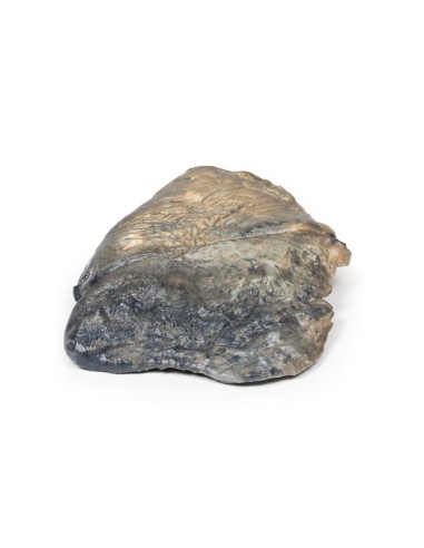
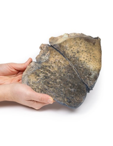
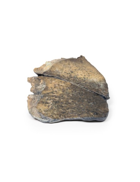
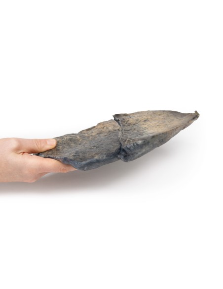
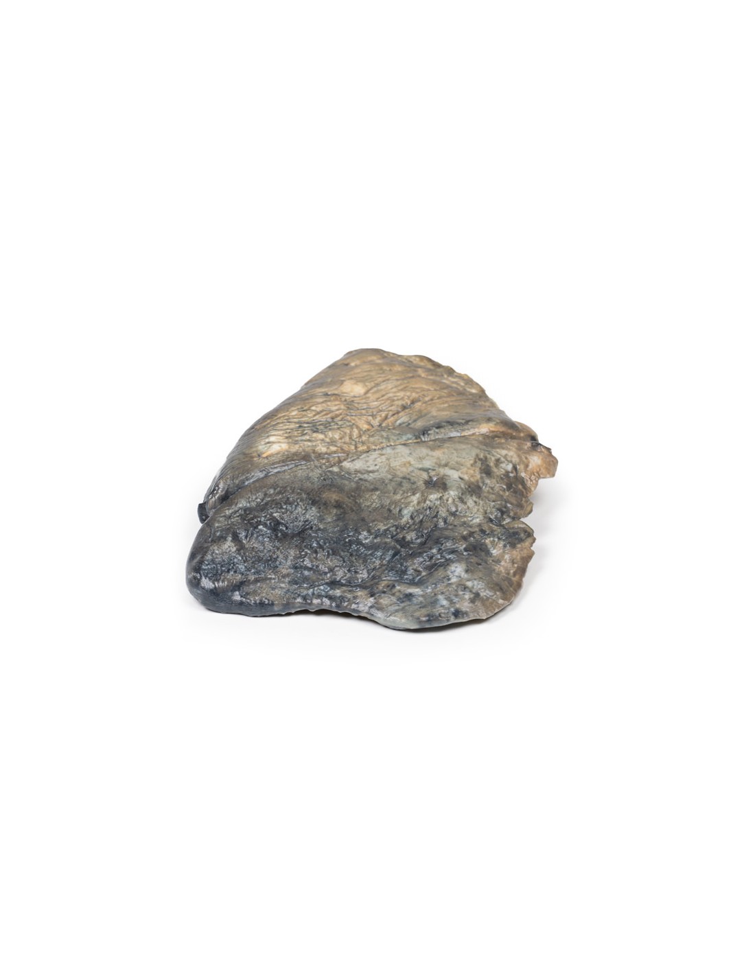
This dissection model highlighting Bronchopneumonia is part of the exclusive Monash 3D anatomy series, a comprehensive series of human dissections reproduced with ultra-high resolution color 3D printing.
Clinical history
There is no clinical history for this specimen.
Pathology
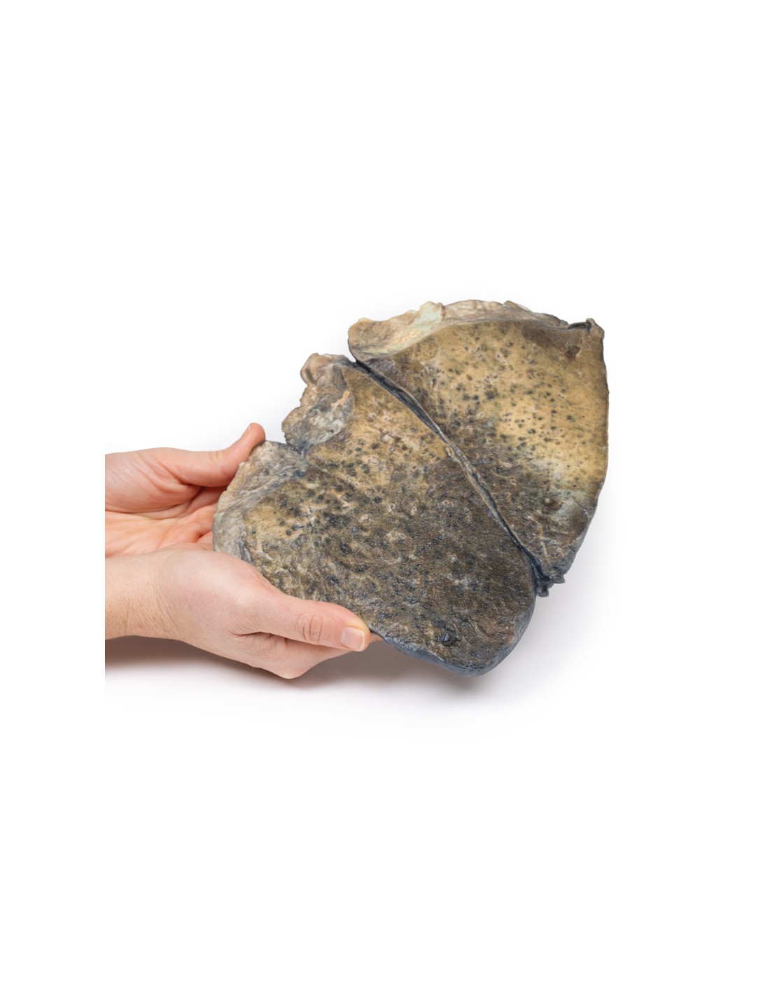
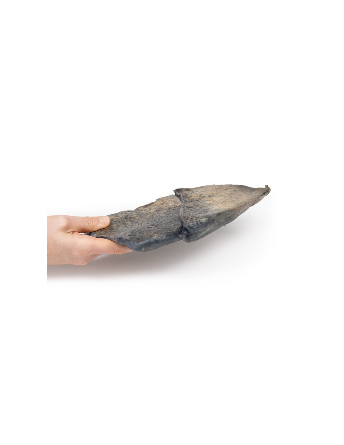
The specimen is a parasagittal section of the left lung. There are irregular regions of focal consolidation and discoloration caused by congested and hyperemic lung tissue distributed within both lobes; however, the upper lobe is more severely affected. Consolidation appears to be concentrated around the bronchioles, which are ectasic. The costal (pleural) surface of the upper lobe is particularly discolored.
Further information
Bronchopneumonia is a form of pneumonia characterized by inflammatory exudate within the intra-alveolar space resulting in consolidation affecting a large, continuous area of the lobe of a lung. It is one of two anatomic classifications of pneumonia (the other is lobar pneumonia). The affected regions in this case show a classic focal red hepatization or consolidation in the focal regions, which is due to vascular congestion with extravasation of red blood cells into the alveolar spaces, along with increased numbers of neutrophils and fibrin. The filling of the airspaces by exudation leads to a gross appearance of solidification, or consolidation, of the alveolar parenchyma.
Bronchopneumonia is a subtype of pneumonia. It is the acute inflammation of the bronchi, accompanied by inflamed patches in the peribronchial and peribronchiolar lobules of the lungs.
It is often contrasted with lobar pneumonia, but in clinical practice the types are difficult to apply because the patterns usually overlap. Bronchopneumonia (sometimes called lobular) often leads to lobar pneumonia as the infection progresses to affect an entire lobe. The same organism can cause one type of pneumonia in one patient and another in another patient. Causes
Bronchopneumonia is usually a bacterial pneumonia rather than caused by a viral disease and is more commonly a hospital-acquired pneumonia than a community-acquired pneumonia, in contrast to lobar pneumonia.
What advantages does the Monash University anatomical dissection collection offer over plastic models or plastinated human specimens?
- Each body replica has been carefully created from selected patient X-ray data or human cadaver specimens selected by a highly trained team of anatomists at the Monash University Center for Human Anatomy Education to illustrate a range of clinically important areas of anatomy with a quality and fidelity that cannot be achieved with conventional anatomical models-this is real anatomy, not stylized anatomy.
- Each body replica has been rigorously checked by a team of highly trained anatomists at the Center for Human Anatomy Education, Monash University, to ensure the anatomical accuracy of the final product.
- The body replicas are not real human tissue and therefore not subject to any barriers of transportation, import, or use in educational facilities that do not hold an anatomy license. The Monash 3D Anatomy dissection series avoids these and other ethical issues that are raised when dealing with plastinated human remains.
No reviews
Tap to zoom
