Your cart
There are no more items in your cart


Metastatic tumor in the lung from primary testicular cancer - Erler Zimmer 3D anatomy Series MP2055
erler zimmer
EZ-MP2055
€921.95
Tax included
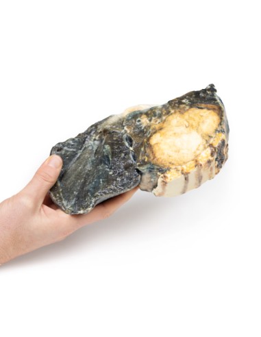
Made in ultra-high resolution 3D printing in full color.
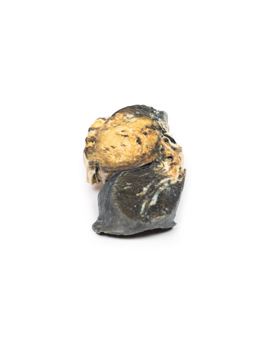
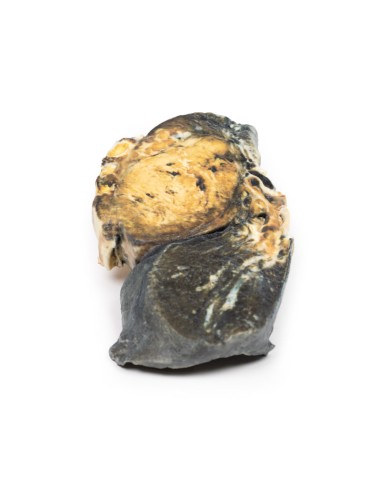
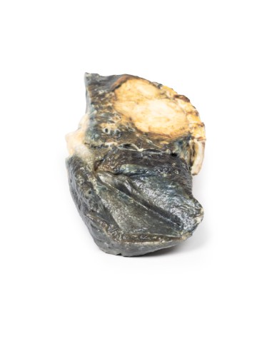
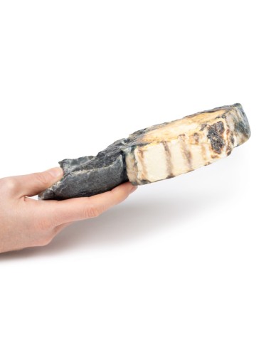
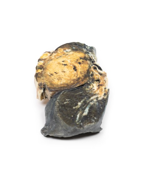
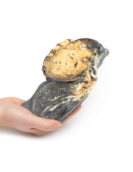
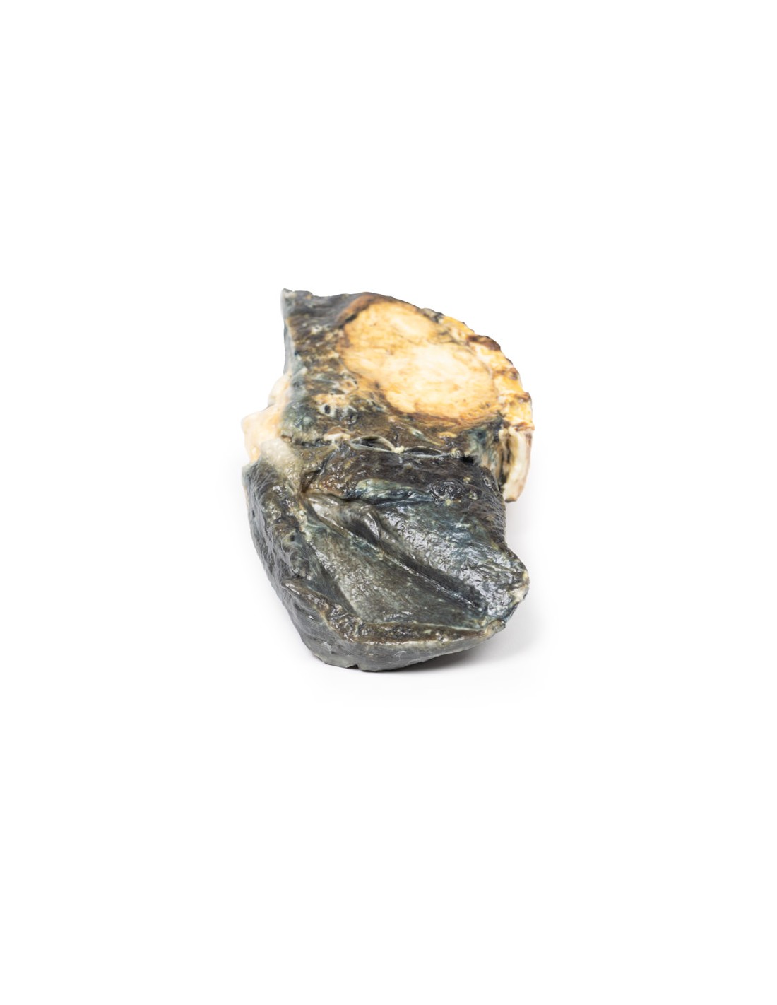
Metastatic Tumor in the Lung from Primary Testicular Cancer - Erler Zimmer 3D anatomy Series MP2055
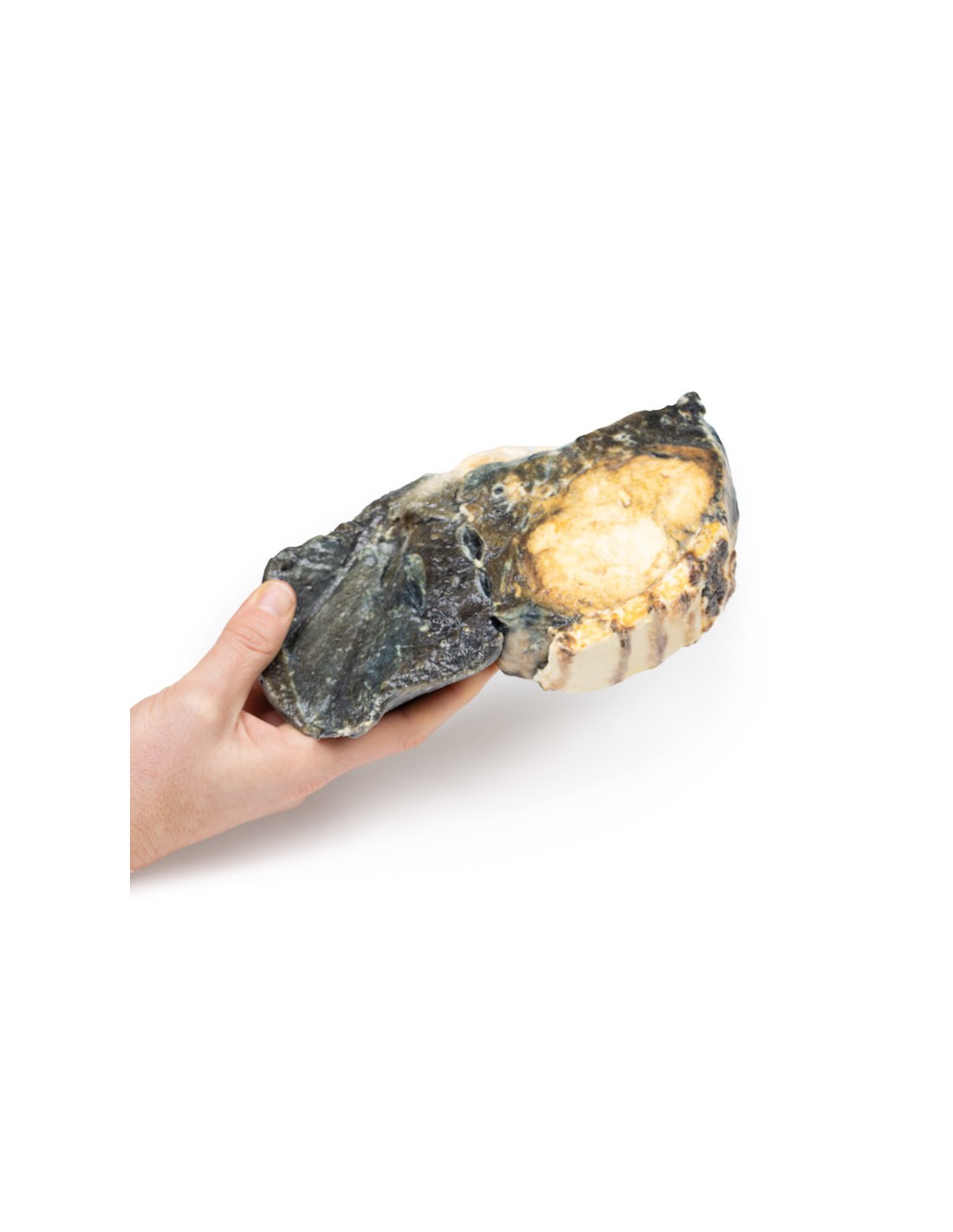
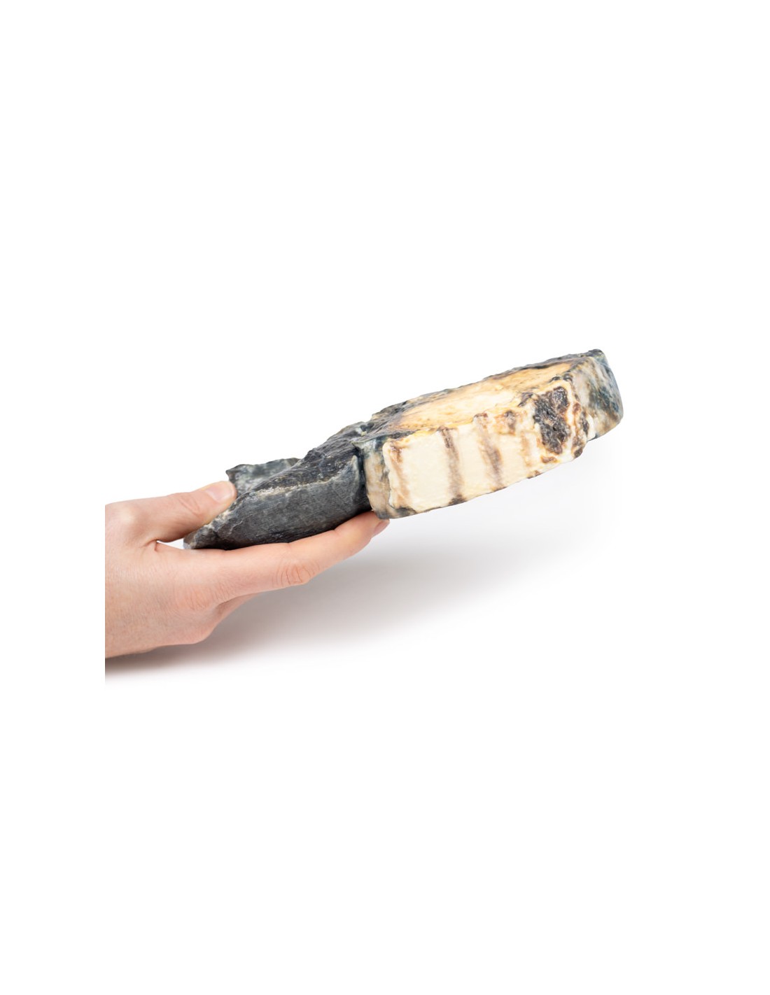
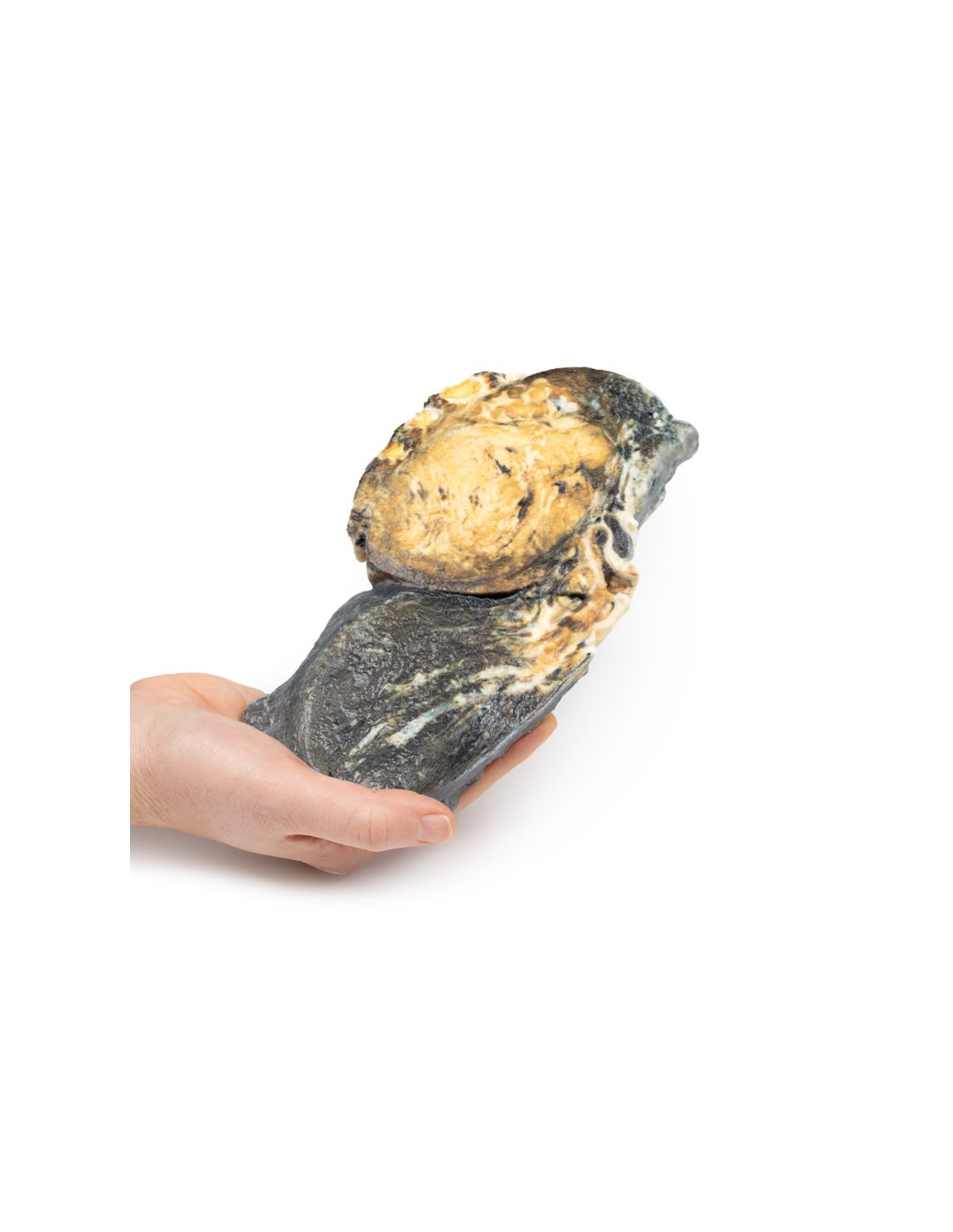
This dissection model highlighting a Metastatic Tumor in the Lung is part of the exclusive Monash 3D anatomy series, a comprehensive series of human dissections reproduced with ultra-high resolution color 3D printing.
Clinical History.
A 37-year-old male patient presented with a 1-month history of lethargy, cough, and weight loss. He had a history of an orchiectomy 18 months earlier for testicular cancer. So 12 months after surgery, he underwent neck radiation therapy to treat metastases. Upon admission, he became acutely dyspnoic and hypoxic and died.
Pathology
This specimen of right lung (and portions of 4 ribs) was sliced longitudinally. There are numerous rounded tumor nodules evident in the lung parenchyma ranging from 5 to 30 mm in diameter. The tumors have a variegated appearance with pale yellow and dark brown cut surfaces. One tumor extends along the lower lobe bronchus forming a cast. Several nodes protrude from the pleural surface and some show a central umbilicus from necrosis and hemorrhage. This is an example of lung metastasis from a testicular mixed germ cell tumor, most likely choriocarcinoma arising in a malignant teratoma.
Further Information.
Testicular germ cell tumors (GCTs) are the most common tumors found in men. The average age of diagnosis is 30 years and they are rarely diagnosed before puberty. Risk factors for development include cryptorchidism and a positive family history of GCT. Increased familial GCT risk may be related to genes coding for kinases, for example KIT and BAK.
They can be divided into two groups: seminomatous (resembling primordial germ cells) and nonseminomatous (resembling embryonic stem cells). More than one-third of GCTs are mixed GCTs, with two or more types of GCTs in a mass. Many possible combinations of seminoma, teratoma, embryonal carcinoma, yolk sac tumor and choriocarcinoma can be observed. Teratoma components are found in one-third of the mixed GCT. High serum levels of alpha fetoprotein and beta-hCG are produced by choriocarcinoma. Lymphatic spread initially involves the retroperitoneal para-aortic lymph nodes. Later mediastinal and supraclavicular lymph nodes may be involved. The lungs are the most common site of hematogenous spread, but the liver, brain, or bones may also be affected.
Symptoms may include a painless testicular mass and hematospermia. Subsequent symptoms of distant metastasis may occur. Common symptoms of lung metastases include cough, dyspnea, hemoptysis, recurrent infections
Treatment depends on the clinical stage but usually involves radical orchiectomy, chemotherapy, and sometimes radiation therapy. More than 95% of early stage GCT can be cured.
What advantages does the Monash University anatomical dissection collection offer over plastic models or plastinated human specimens?
- Each body replica has been carefully created from selected patient X-ray data or human cadaver specimens selected by a highly trained team of anatomists at the Monash University Center for Human Anatomy Education to illustrate a range of clinically important areas of anatomy with a quality and fidelity that cannot be achieved with conventional anatomical models-this is real anatomy, not stylized anatomy.
- Each body replica has been rigorously checked by a team of highly trained anatomists at the Center for Human Anatomy Education, Monash University, to ensure the anatomical accuracy of the final product.
- The body replicas are not real human tissue and therefore not subject to any barriers of transportation, import, or use in educational facilities that do not hold an anatomy license. The Monash 3D Anatomy dissection series avoids these and other ethical issues that are raised when dealing with plastinated human remains.
No reviews
Tap to zoom