Your cart
There are no more items in your cart


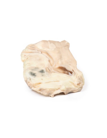
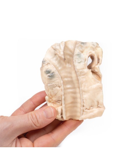
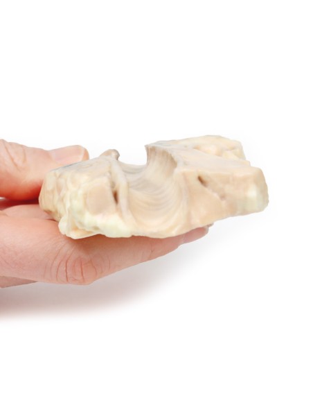
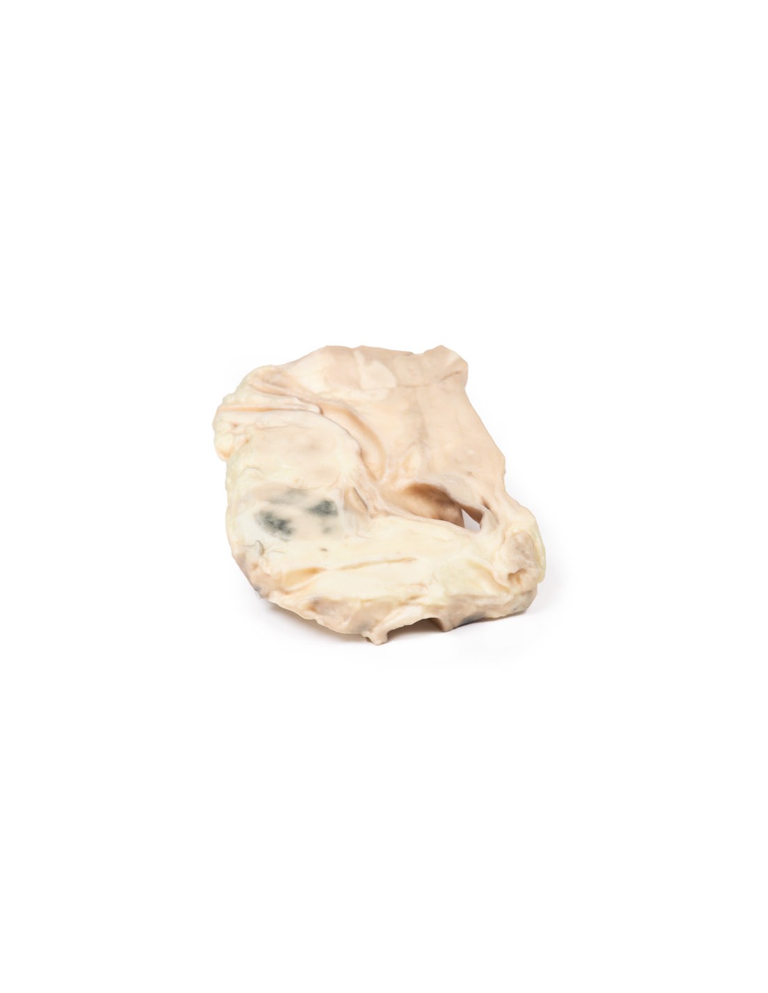
Trachea: Hodgkin's lymphoma- Erler Zimmer 3D anatomy Series MP2062
erler zimmer
EZ-MP2062
€322.08
Tax included
Made in ultra-high resolution 3D printing in full color.
Trachea: Hodgkin's lymphoma- Erler Zimmer 3D anatomy Series MP2062
This dissection model highlighting a Trachea: Hodgkin's lymphoma is part of the exclusive Monash 3D anatomy series, a comprehensive series of human dissections reproduced with ultra-high resolution color 3D printing.
Clinical History.
A 45-year-old man presented with a nodule in his left supraclavicular area. The swelling had increased in size over 6 months. Biopsy excision of the nodule showed Hodgkin's lymphoma (HL). Ten months later, he was readmitted with left shoulder pain and left arm swelling. Examination revealed generalized lymphadenopathy with significant swelling in the left supraclavicular and axillary regions. He was treated with radiotherapy and Thiotepa chemotherapy. He developed vomiting. A subsequent barium meal showed duodenal obstruction from extrinsic lymph node compression. He continued to deteriorate and died 2 weeks after readmission.
Pathology
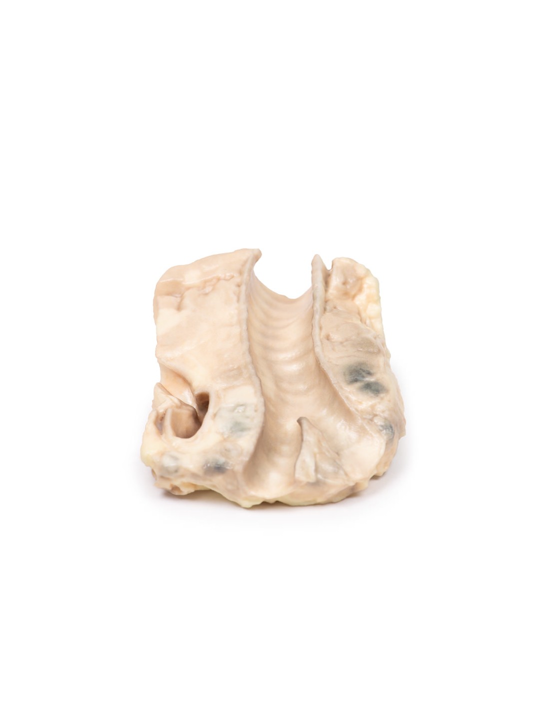
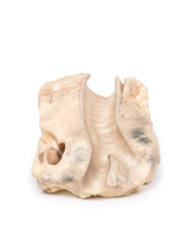
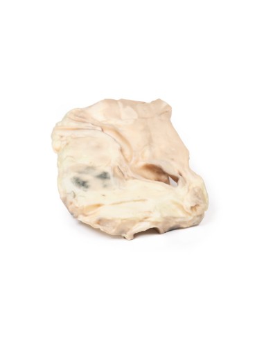
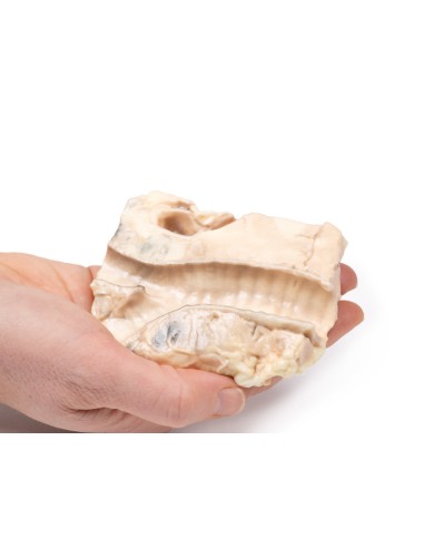
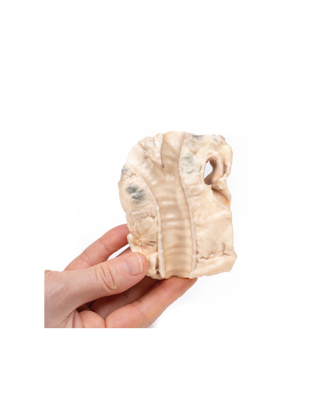
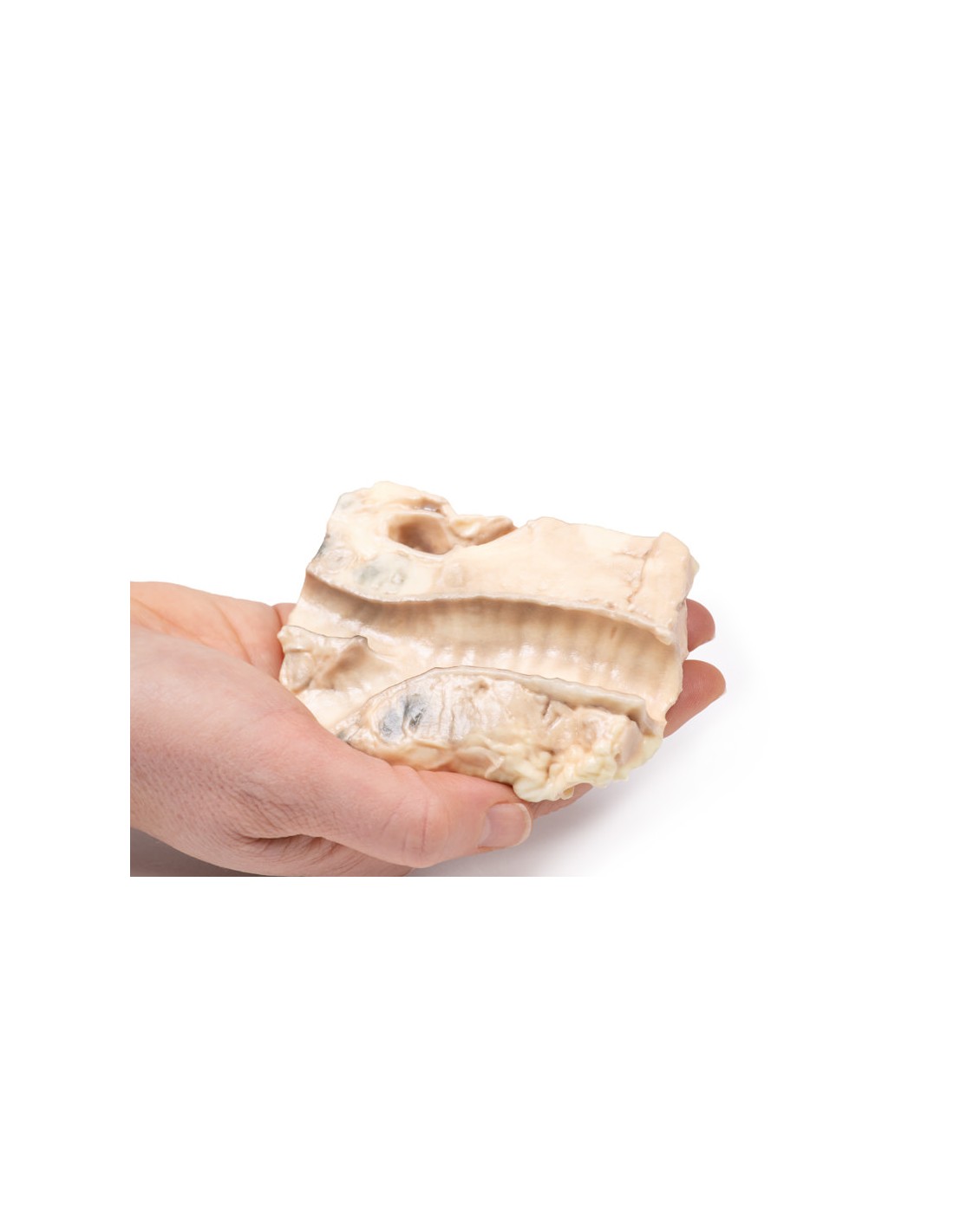
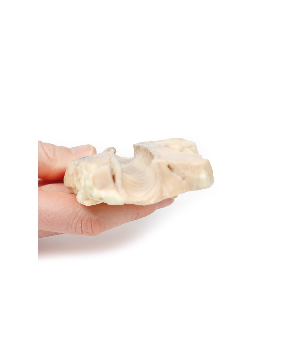
3D print shows tracheal bifurcation with adjacent para-tracheal and peri-bronchial lymph nodes. The trachea has been opened longitudinally and is seen from behind. The para-tracheal lymph nodes are pale and ruffled (fused) together. Similar abnormal tissue is seen as a confluent pale mass on the left side of the trachea, above the aortic arch, which is seen cut in cross section as a hollow space with branching. The peribronchial lymph nodes are also enlarged and contain carbon pigment. Smaller lighter circumscribed areas in the lymph nodes and extra-nodal tumor are foci of necrosis. There is an atheroma in the wall of the aorta but it is difficult to see in 3D printing.
More information
Hodgkin's lymphoma (HL) is a malignant tumor of lymphocytes. It is characterized by the presence of neoplastic giant cells called Reed Sternberg cells. There are 5 major subtypes according to the WHO classification of lymphomas, based on morphology, immunophenotyping, and genetics. Activation of the transcription factor NF-kB is a common pathway of tumorigenesis among the subtypes. This promotes proliferation, reduces apoptosis, and induces the expression of cytokines that recruit immune cells surrounding Reed Sternberg cells in HL.
There is a bimodal distribution by age with a peak in late adolescence/early adulthood and a second peak in the elderly. HL accounts for slightly less than 1% of all cancers worldwide. Epstein Barr Virus (EBV) infection contributes to the pathogenesis of major HL subtypes. The viral genome causes genetic alterations that lead to aberrant signaling pathways, although the precise mechanism is not known. Immunosuppression (e.g., HIV infection or organ transplantation) and positive family history for HL are also risk factors. HL commonly presents as painless lymphadenopathy, itching, weight loss, fevers, and night sweats. Later the disease sees the organ spread to the spleen, liver, and bone marrow. Compressive symptoms may result from enlarged lymph nodes and infiltrated organs. HL is diagnosed by staging CT scan, biopsy for excision of involved lymph nodes, and bone marrow biopsy. Treatment involves radiotherapy and chemotherapy. Although previously incurable, the overall survival of HL has greatly improved over the past 5 decades with modern therapies: diagnosed at an early stage, it is nearly 90%, and even advanced disease has a favorable prognosis.
What advantages does the Monash University anatomical dissection collection offer over plastic models or plastinated human specimens?
- Each body replica has been carefully created from selected patient X-ray data or human cadaver specimens selected by a highly trained team of anatomists at the Monash University Center for Human Anatomy Education to illustrate a range of clinically important areas of anatomy with a quality and fidelity that cannot be achieved with conventional anatomical models-this is real anatomy, not stylized anatomy.
- Each body replica has been rigorously checked by a team of highly trained anatomists at the Center for Human Anatomy Education, Monash University, to ensure the anatomical accuracy of the final product.
- The body replicas are not real human tissue and therefore not subject to any barriers of transportation, import, or use in educational facilities that do not hold an anatomy license. The Monash 3D Anatomy dissection series avoids these and other ethical issues that are raised when dealing with plastinated human remains.
No reviews
Tap to zoom