Your cart
There are no more items in your cart


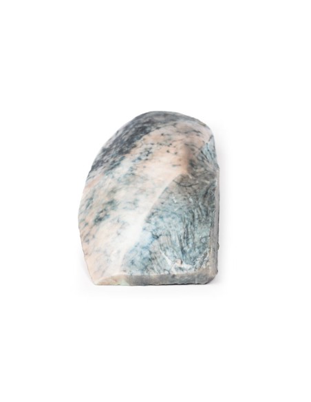
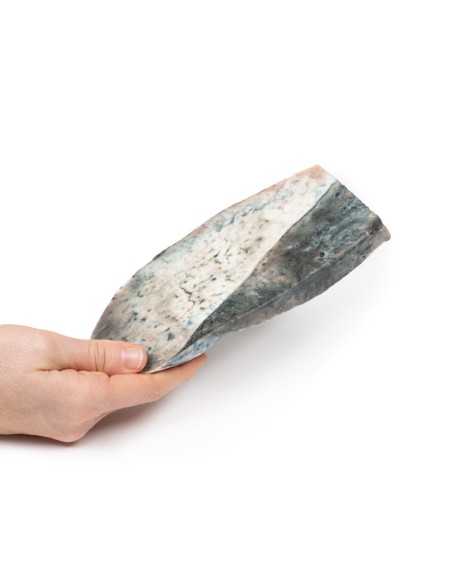
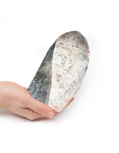
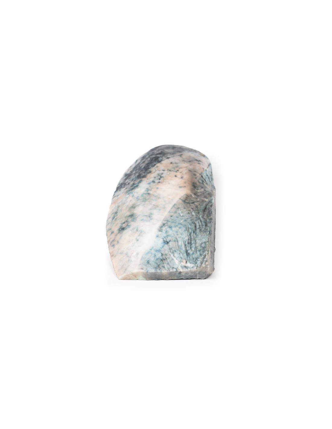
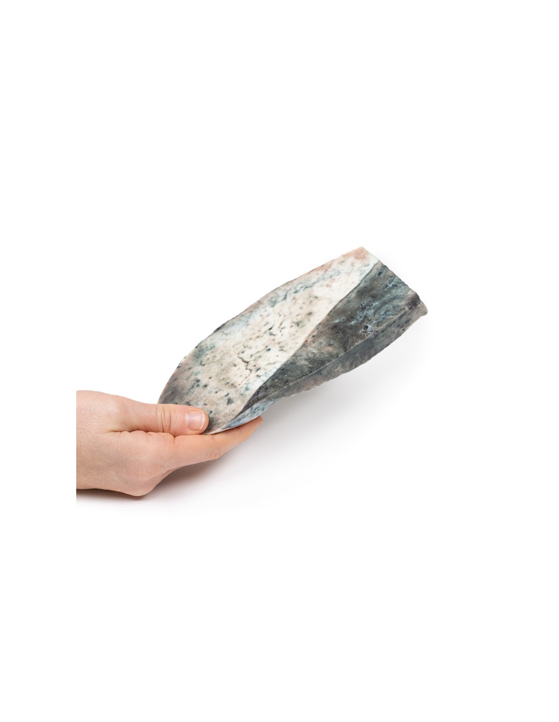
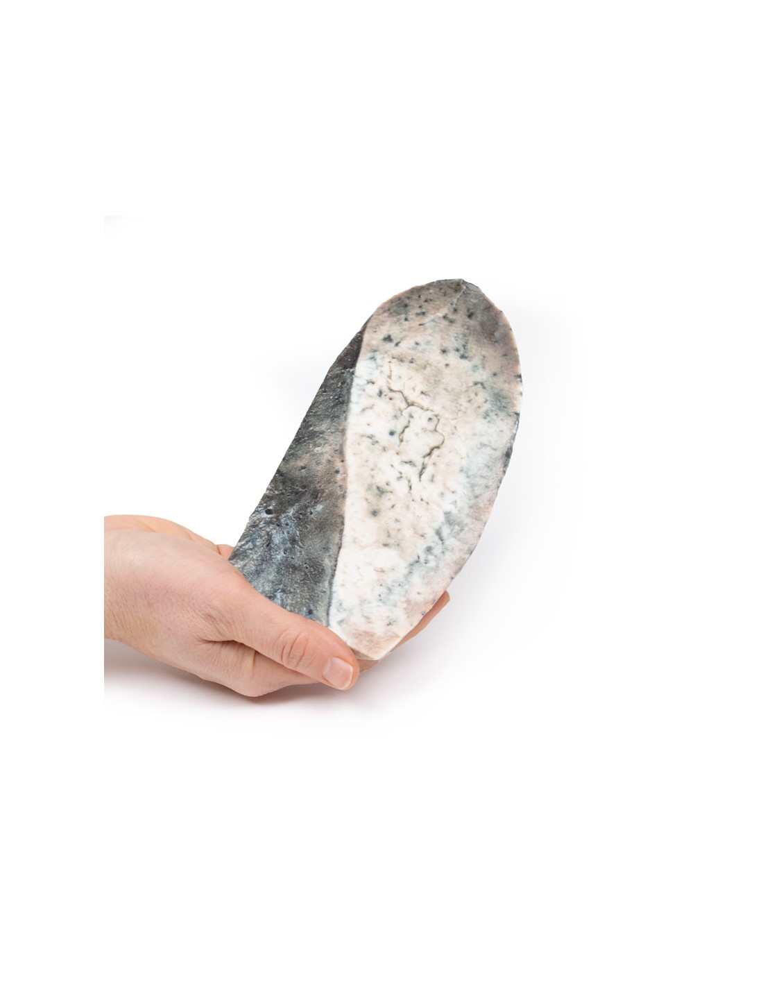
Lobar pneumonia - Gray hepatization phase - Erler Zimmer 3D anatomy Series MP2061
erler zimmer
EZ-MP2061
€446.89
Tax included
Made in ultra-high resolution 3D printing in full color.
Lobar pneumonia - Gray hepatization phase - Erler Zimmer 3D anatomy Series MP2061
This dissection model highlighting a Lobar Pneumonia - Gray Hepatization Phase is part of the exclusive Monash 3D anatomy series, a comprehensive series of human dissections reproduced with ultra-high resolution color 3D printing.
Clinical history
There is no clinical history for this specimen.
Pathology
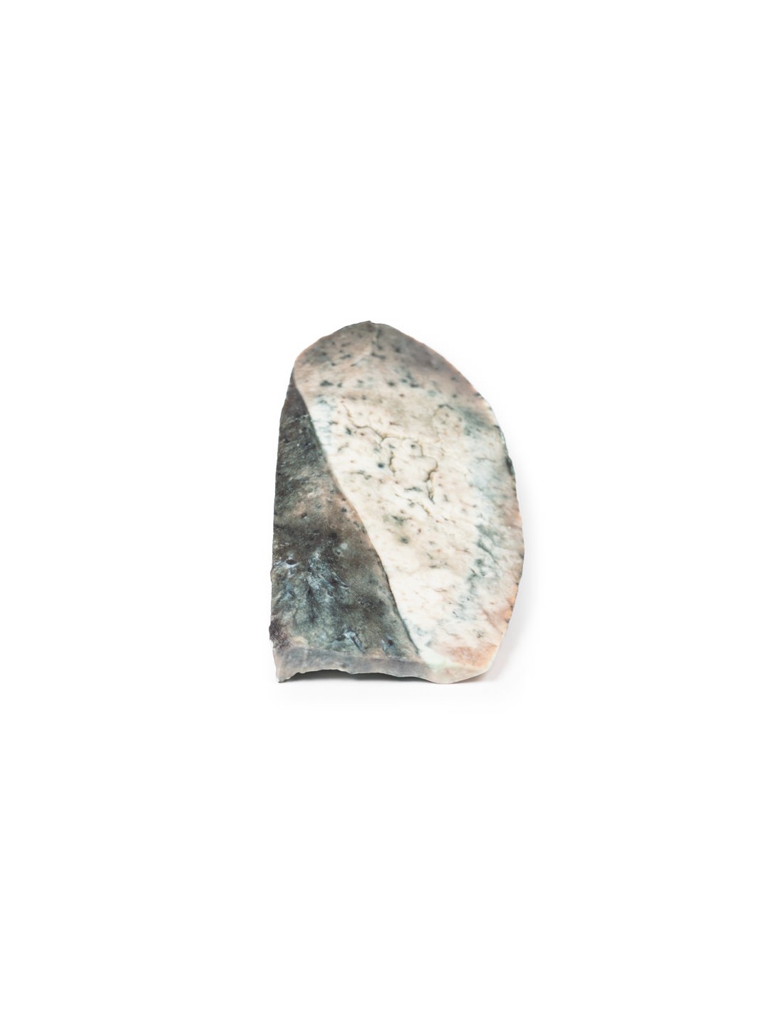
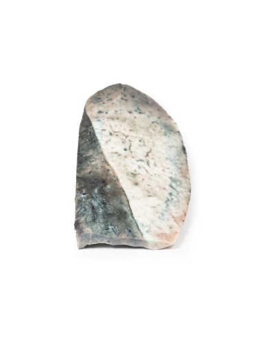
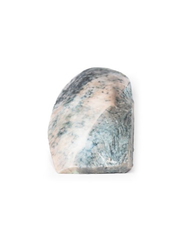
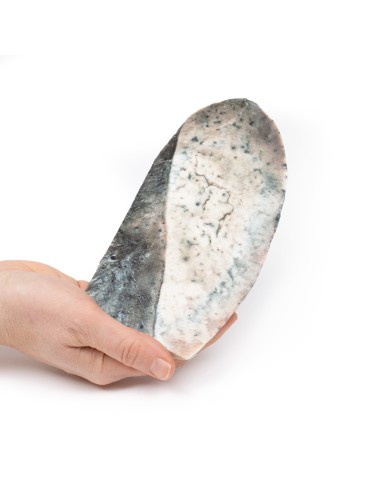
The specimen is a parasagittal section of the right lung, and the boundary between the upper and lower lobes is clearly visible. The entire upper lobe is congested and light gray in color.
Additional information
This is an example of a stage of lobar pneumonia in which inflammatory exudates within the intra-alveolar space result in consolidation affecting a large and continuous area of the lobe of one lung. The affected lobe in this case shows gray hepatization or late consolidation. This usually occurs 2 to 3 days after red hepatization and lasts 4 to 8 days. The lung appears gray with solid liver-like consistency due to a fibrinopurulent exudate, progressive disintegration of red blood cells, and hemosiderin. Large numbers of macrophages begin to appear in the interstitial tissue. They are the dominant cells that attempt to clear cellular debris and acute inflammation through phagocytosis. Macrophages may contain iron due to erythrocyte consumption and are therefore called siderophages. After gray hepatization, resolution and restoration of lung architecture begin by day 8. Enzymatic action begins centrally and spreads peripherally, which liquefies the previous fibrinous solid content and eventually restores aeration.
The most common organisms causing lobar pneumonia are Streptococcus pneumoniae, also called pneumococcus, Haemophilus influenzae, and Moraxella catarrhalis. Mycobacterium tuberculosis, the tubercle bacillus, can also cause lobar pneumonia if pulmonary tuberculosis is not treated promptly. Other microorganisms that cause lobar pneumonia are Legionella pneumophila and Klebsiella pneumoniae.[2]In a posterioanterior and lateral chest radiograph, an entire lobe will be radiopaque, which is indicative of lobar pneumonia.
What advantages does the Monash University anatomical dissection collection offer over plastic models or plastinated human specimens?
- Each body replica has been carefully created from selected patient X-ray data or human cadaver specimens selected by a highly trained team of anatomists at the Monash University Center for Human Anatomy Education to illustrate a range of clinically important areas of anatomy with a quality and fidelity that cannot be achieved with conventional anatomical models-this is real anatomy, not stylized anatomy.
- Each body replica has been rigorously checked by a team of highly trained anatomists at the Center for Human Anatomy Education, Monash University, to ensure the anatomical accuracy of the final product.
- The body replicas are not real human tissue and therefore not subject to any barriers of transportation, import, or use in educational facilities that do not hold an anatomy license. The Monash 3D Anatomy dissection series avoids these and other ethical issues that are raised when dealing with plastinated human remains.
No reviews
Tap to zoom