Your cart
There are no more items in your cart


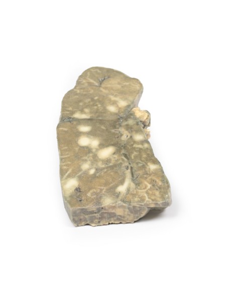
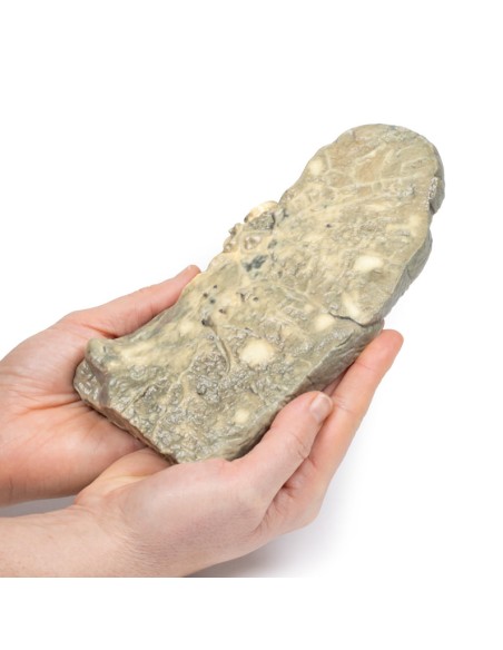
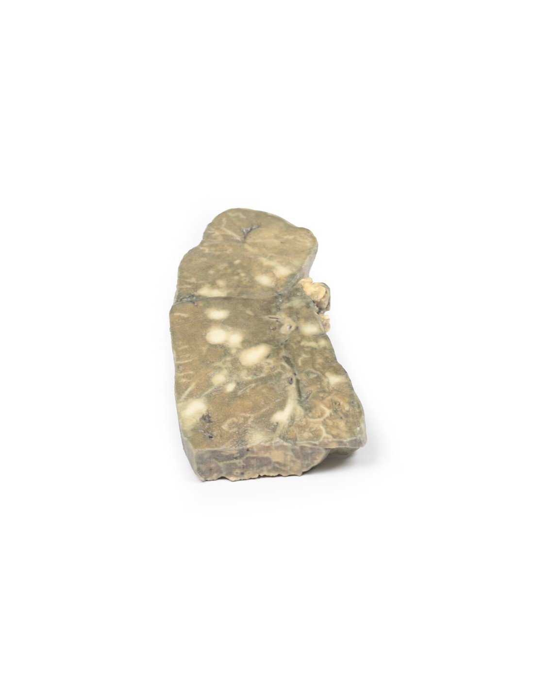
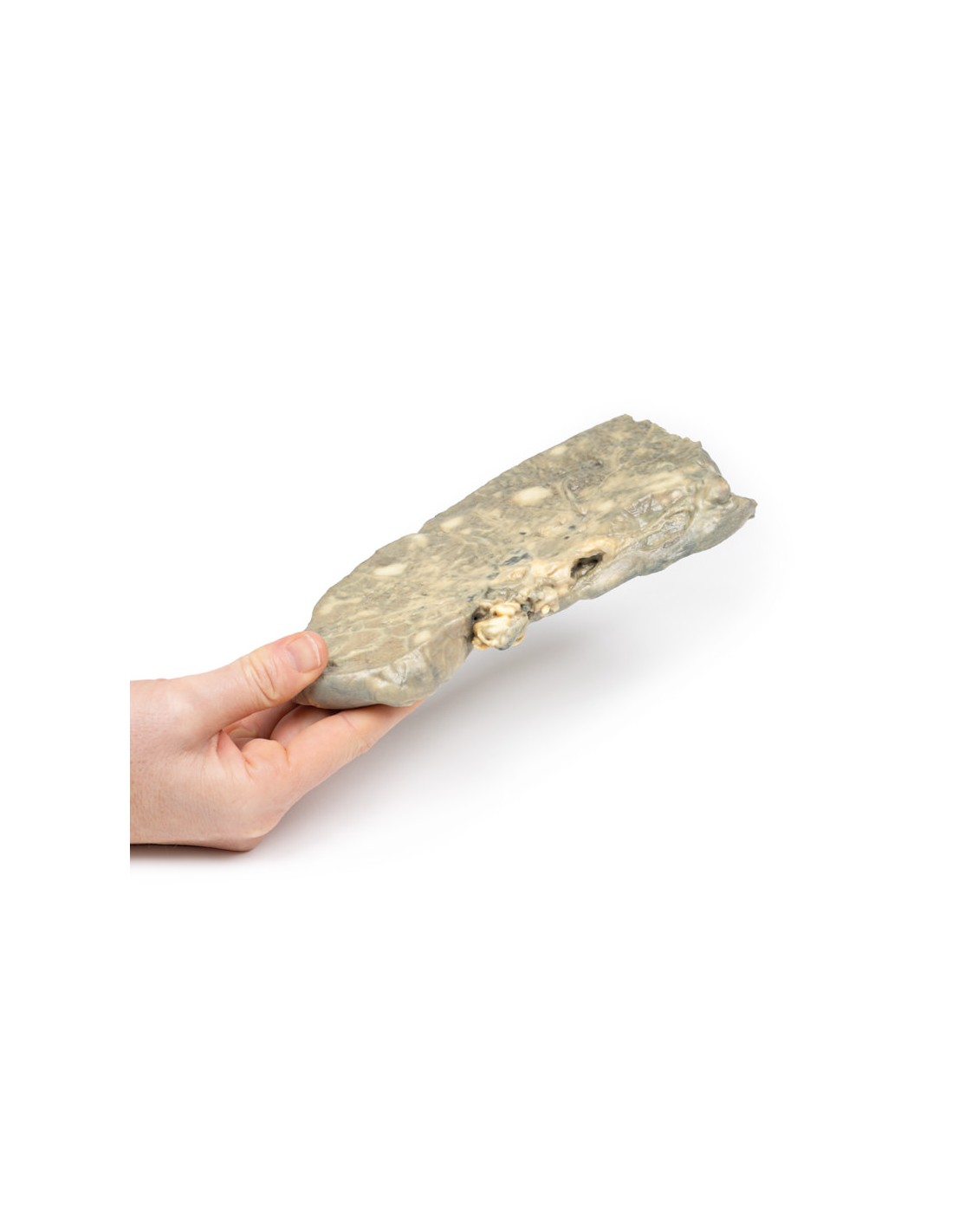
Metastatic carcinoma in the lung - Erler Zimmer 3D anatomy Series MP2056
erler zimmer
EZ-MP2056
€522.04
Tax included
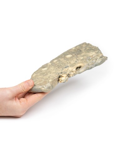
Made in ultra-high resolution 3D printing in full color.
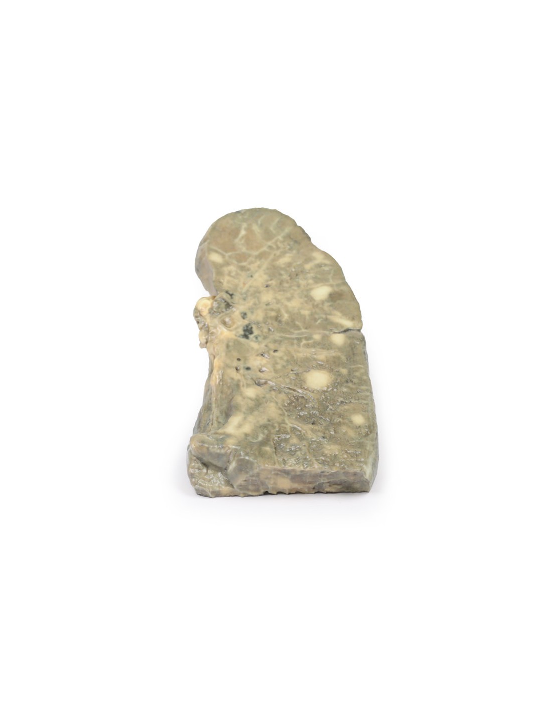
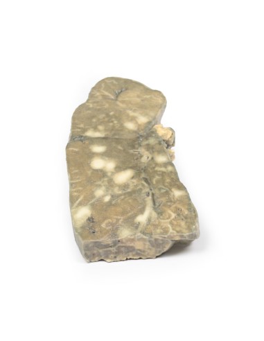
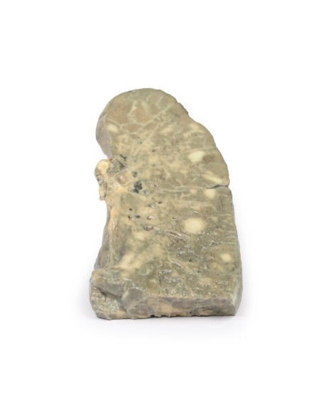
Metastatic Carcinoma in the Lung - Erler Zimmer 3D anatomy Series MP2056
This dissection model highlighting Metastatic Carcinoma in the Lung is part of the exclusive Monash 3D anatomy series, a comprehensive series of human dissections reproduced with very high resolution color 3D printing.
Medical History.
This 47-year-old woman was admitted for terminal carcinomatosis. On examination, a hard liver and a right pelvic mass were palpable. She had been ill with constitutional symptoms for months and eventually sought medical care. She was admitted for palliative care and died shortly thereafter.
Pathology
The left lung was sliced longitudinally to visualize the cut surface. Multiple pale tumor nodules of variable size are scattered throughout the lung substance. Several nodules converge near the hilum. Hilar lymph nodes contain pale tumor tissue. Small tumor nodules can be seen under the thickened pleura. Histologically, these were metastatic deposits of adenocarcinoma. At autopsy, there was adenocarcinoma of the ovary, with metastasis to the lungs, heart, liver, and pericardium.
Further information
Lung metastases are more common than primary lung carcinoma. Malignant disease arising anywhere in the body can spread to the lungs. Sarcomas usually metastasize into the bloodstream, and carcinomas spread via the bloodstream or the lymphatic system or both.
What advantages does the Monash University anatomical dissection collection offer over plastic models or plastinated human specimens?
- Each body replica has been carefully created from selected patient X-ray data or human cadaver specimens selected by a highly trained team of anatomists at the Monash University Center for Human Anatomy Education to illustrate a range of clinically important areas of anatomy with a quality and fidelity that cannot be achieved with conventional anatomical models-this is real anatomy, not stylized anatomy.
- Each body replica has been rigorously checked by a team of highly trained anatomists at the Center for Human Anatomy Education, Monash University, to ensure the anatomical accuracy of the final product.
- The body replicas are not real human tissue and therefore not subject to any barriers of transportation, import, or use in educational facilities that do not hold an anatomy license. The Monash 3D Anatomy dissection series avoids these and other ethical issues that are raised when dealing with plastinated human remains.
No reviews
Tap to zoom