Your cart
There are no more items in your cart


Made in ultra-high resolution 3D printing in full color.
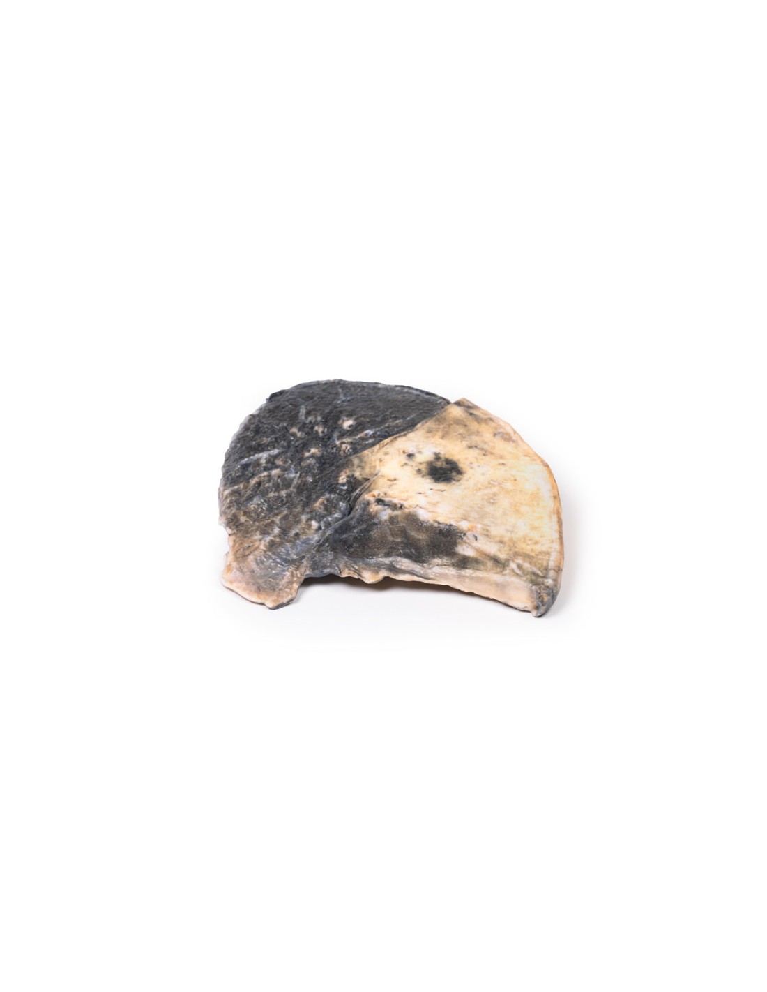
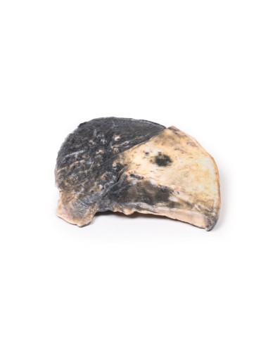
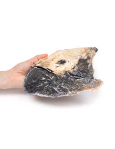
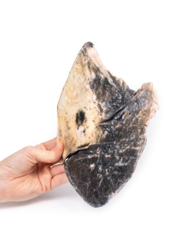
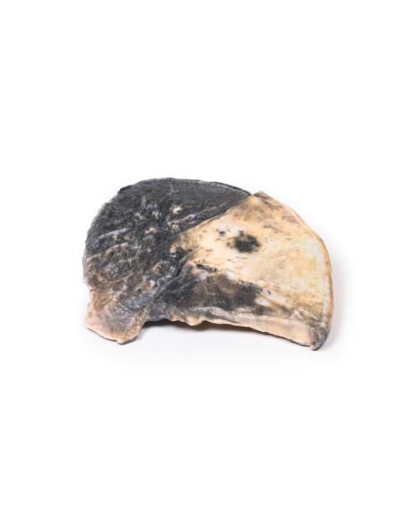
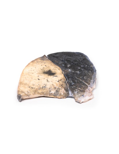
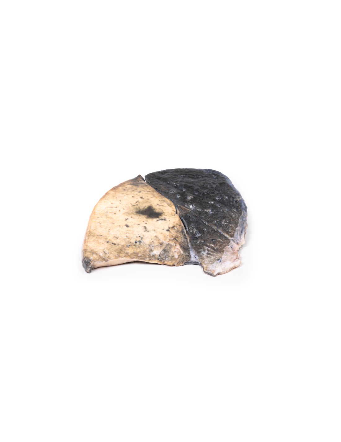
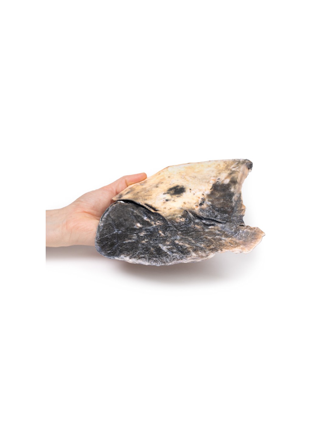
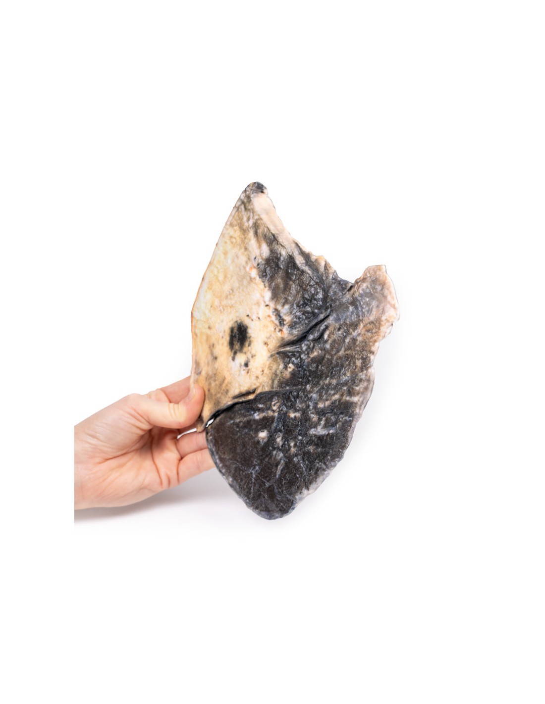
Lobar pneumonia - Erler Zimmer 3D anatomy Series MP2057
This dissection model highlighting lobar pneumonia is part of the exclusive Monash 3D anatomy series, a comprehensive series of human dissections reproduced with ultra-high resolution color 3D printing.
Clinical history
There is no clinical history for this specimen.
Pathology
The specimen is a parasagittal section of the right lung, and the boundaries between the three lobes are visible. The entire upper and middle lobes are congested and hyperemic* causing the darker appearance. There are smaller foci in the left lung.
Further information
Lobar pneumonia is a form of pneumonia characterized by inflammatory exudate within the intra-alveolar space with subsequent consolidation affecting a large, continuous area of the lobe of a lung. It is one of two anatomical classifications of pneumonia (the other is bronchopneumonia). The affected lobe in this case shows the classic red "hepatization" or consolidation of the lung parenchyma, which is due to vascular congestion with overflow of red blood cells into the alveolar spaces, along with increased numbers of neutrophils and fibrin. The filling of the air spaces by exudation leads to a coarse appearance of solidification, or consolidation, of the alveolar parenchyma. This reddish appearance has been compared to that of the cut surface of the liver, hence the term "hepatization."
The most common organisms causing lobar pneumonia are Streptococcus pneumoniae, also called pneumococcus, Haemophilus influenzae, and Moraxella catarrhalis. Mycobacterium tuberculosis, the tubercle bacillus, can also cause lobar pneumonia if pulmonary tuberculosis is not treated promptly. Other organisms that lead to lobar pneumonia are Legionella pneumophila and Klebsiella pneumoniae.
Like other types of pneumonia, lobar pneumonia can occur as a community-acquired infection, in immunosuppressed patients, or as a nosocomial infection. However, most of the causative organisms are of the community-acquired type.
On a posteroanterior and lateral chest radiograph, an entire lobe will be radiopaque without evidence of air within it, indicative of lobar pneumonia.
*Hypermia = active engorgement of the vascular beds with normal or reduced blood flow.
What advantages does the Monash University anatomical dissection collection offer over plastic models or plastinated human specimens?
- Each body replica has been carefully created from selected patient X-ray data or human cadaver specimens selected by a highly trained team of anatomists at the Monash University Center for Human Anatomy Education to illustrate a range of clinically important areas of anatomy with a quality and fidelity that cannot be achieved with conventional anatomical models-this is real anatomy, not stylized anatomy.
- Each body replica has been rigorously checked by a team of highly trained anatomists at the Center for Human Anatomy Education, Monash University, to ensure the anatomical accuracy of the final product.
- The body replicas are not real human tissue and therefore not subject to any barriers of transportation, import, or use in educational facilities that do not hold an anatomy license. The Monash 3D Anatomy dissection series avoids these and other ethical issues that are raised when dealing with plastinated human remains.
No reviews
Tap to zoom