- -15%
Femur fracture and hip dislocation 3B Scientific A88
3b scientificFemur fracture and hip dislocation A88
Anatomical models 3B scientific, femur fracture and hip dislocation A88:
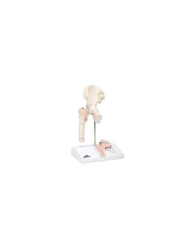
Anatomical model of femur fracture and hip dislocation.
This 3B Scientific anatomical model serves as an aid to give understandable explanations to patients, for example, before operations.
It shows the right hip of a man of a certain age, half the natural size. On the base also there is a frontal section of the femoral neck in relief.
The model shows the most common femur fractures in practice and typical forms of hip dislocation (coxarthrosis or hip arthrosis). The following fractures are illustrated:
- middle fracture of the femoral neck
- lateral fracture of the femoral neck
- transverse fracture of the trochanter
- fracture under the trochanter
- fracture in the tubular area of the femur
- fracture in the head area of the femur
- fracture of the greater trochanter
- Fracture or tear of the small trochanter.
On a removable basis
The 3B Scientific A88 anatomical femur fracture and hip dislocation model is made of polyvinyl chloride (PVC), which is very hard, lightweight, and corrosion resistant. PVC resists reactions with acids, alcohol gasoline, and hydrocarbons.
Pleasenote, item mounted on removable base included in package
Why buy it
3B Scientific Anatomical Models are undoubtedly the best on the market, ideal for teaching, providing clarification to patients, and scientific medical training.
Many physicians and professionals purchase the anatomical teaching models to highlight key points on topics such as the skeleton, human musculature, joints and related diseases (rheumatism, arthrosis, bursitis, synovitis, bursitis, tendonitis, cervical, ischialgia).
They can also be used as furnishing accessories to personalize one's medical office.
- Height
- 22
- Width
- 14
- Depth
- 10
- Weight
- 1


