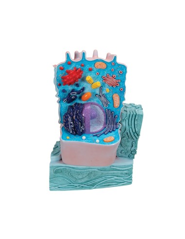
Enlarged by about 10,000 times
Animal cell model - 3B Scientific R04:
Two-part model shows shape and structures of a typical animal cell by observation under an electron microscope. All major organelles are made in relief and represented by color differentiation:
- cell nucleus
- mitochondrion
- smooth endoplasmic reticulum
- rough endoplasmic reticulum
- basement membrane
- collagen fibers
- Golgi apparatus
- microvilli
- lysosome
Why buy it
3B Scientific Anatomical Teaching Models are undoubtedly the best on the market, ideal for teaching, for providing clarification to patients, and for scientific medical training.
Many physicians and professionals purchase the anatomical teaching models to highlight key points on topics such as the skeleton, human musculature, joints and related diseases (rheumatism, arthritis, bursitis, synovitis, bursitis, tendonitis, cervical, ischialgia).
They can also be used as furnishing accessories to personalize one's medical office.
- Height
- 21
- Width
- 11
- Depth
- 31
- Weight
- 1


