Your cart
There are no more items in your cart
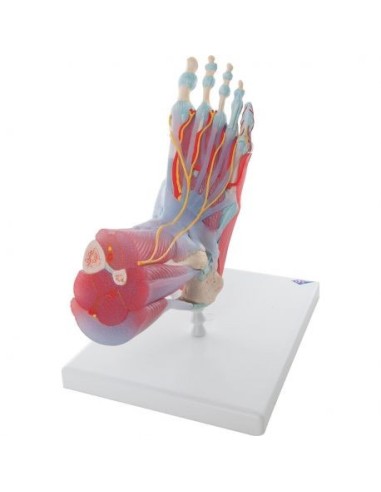

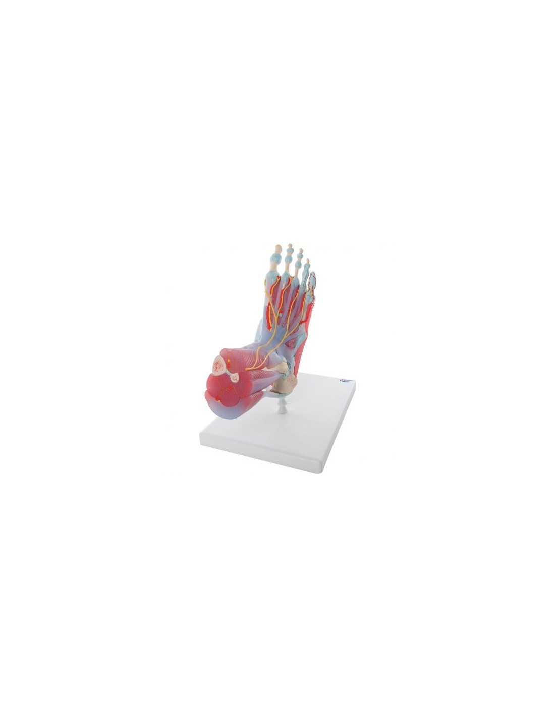
Model of foot skeleton with ligaments and muscles 3B Scientific M34/1
3b scientific
1495
Last items in stock
€347.00
Tax included
Anatomically detailed model of the foot and calf that can be disassembled into 6 removable pieces.
Anatomical model of foot skeleton with ligaments and muscles 3B Scientific M34/1:
Life-sizeanatomical model of the foot skeleton with evidence of bones, tendons, ligaments, and muscles.
This anatomically detailed model of the foot and calf can be disassembled into 6 removable pieces to allow detailed study of the region.
The anatomical model offers not only bones but also muscles, tendons, ligaments, nerves, arteries, and veins.
The front view shows the extensor muscles of the calf. The tendons can be followed as they pass under the transverse and cruciate ligaments of the thigh to their insertion points. All tendon sheaths are also visible.
Features of the anatomical model of the foot skeleton with ligaments and muscles
On the dorsal portion of the model, the gastrocnemius muscle is removable to allow deeper anatomical elements to be observed. The sole of the foot is shown in three layers. The first shows the short flexor of the toes. This muscle can be excised to highlight the square of the plant, the tendon of the flexor longus of the toes, and the flexor hallucis. In turn, this second layer can be excised to highlight deeper anatomical details.
The 3B Scientific M34/1 anatomical teaching model of foot skeleton with ligaments and muscles is made of polyvinyl chloride (PVC), which is very hard, lightweight, and corrosion resistant. PVC resists reactions with acids, alcohol gasoline, and hydrocarbons.
Why buy an anatomical foot skeleton model?
With the anatomical foot skeleton model with ligaments and muscles, you can study even the smallest details of the anatomy of the human foot, and the interactions between the bones, ligaments, and muscles.
You can also use the skeleton of the foot M34/1 to give explanations to your patients, to make focuses on the pathologies that afflict this anatomical structure and the therapier that will be undertaken to solve the patient's problem.
Communication will become extremely effective with the use of an anatomical model of the skeleton of the foot, the ability to visualize directly on the skeleton of the foot the pathology will make the doctor's or physical therapist's explanation much more incisive.
3B Scientific Anatomical Models are undoubtedly the best on the market, ideal for teaching, for providing clarification to patients, and for scientific medical training.
Many physicians and professionals purchase the anatomical teaching models to highlight key points on topics such as the skeleton, human musculature, joints, and related diseases (rheumatism, osteoarthritis, bursitis, synovitis, bursitis, tendonitis, cervical, ischialgia).
They can also be used as furnishing accessories to personalize one's medical office.
- Height
- 23
- Width
- 16
- Depth
- 19
- Weight
- 1,1
No reviews
Tap to zoom