
Your cart
There are no more items in your cart


Erler Zimmer skull decomposable into 22 parts colored 4708
erler zimmer
EZ-4708
€380.00
Tax included
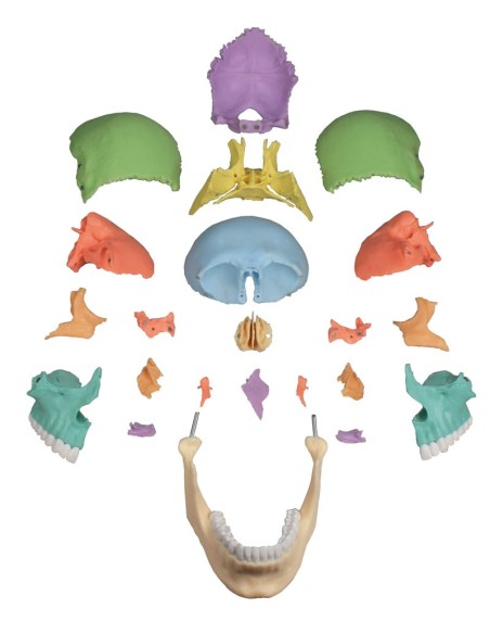
22 Single colored bones
Erler Zimmer skull decomposable into 22 parts colored 4708:
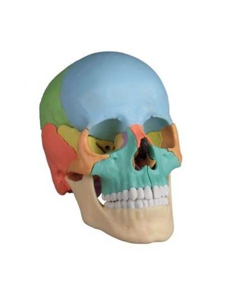
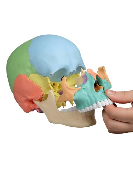
Erler zimmer skull 4708 is a decomposable anatomical model of the human skull, simple to understand, is an excellent choice for basic anatomical study and is also recommended as a medical gift.
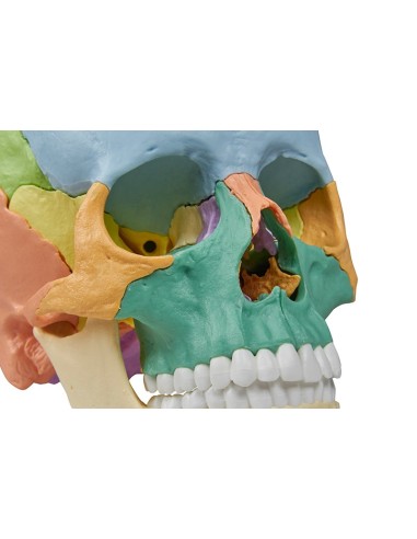
The human skull is composed of many individual bones that adhere to each other only in the course of development. The new Erler Zimmer decomposable skull is a natural cast that clearly highlights the complex structure of the skull, as it can be broken down into 22 bones.
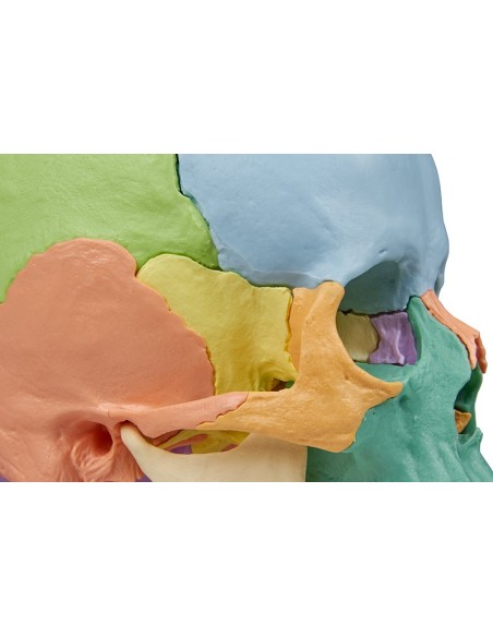
The individual bones can be joined again thanks to the robust and almost invisible connecting elements found in the slightly simplified cranial sutures.
Studying the anatomy of the skull has never been easier; all the bones of the human skull are present in detail in the anatomical model:
- Parietal bone (right and left)
- Occipital bone
- Frontal bone
- Temporal bone (right and left)
- Sphenoid
- Ethmoid
- Vomer
- Zygomatic bone (right and left)
- Upper jaw with teeth (right and left)
- Palatine bone (right and left)
- Nasal cornet (right and left)
- Lacrimal bone (right and left)
- Nasal bone (right and left)
- Lower jaw with teeth
The skull cap is held in line with the skull base by metal pins and held in place by powerful magnets. Because of this, it was not necessary to add plastic pins that could break, and the skull cap adheres perfectly to the skull base.
This detail distinguishes the Erler Zimmer decomposable skull from other anatomical models, with no plastic pins or unsightly external metal joints.
How was the Erler Zimmer skull made?
The Erler Zimmer sk ull represents a unique technological innovation in the field of educational anatomical models.
To make this beautiful educational human skull a real skull was 3d scanned and underwent a digital modeling stage to insert the magnets, slightly simplifying the sutures between its component bones.
The next stage of production was done with very high-resolution 3D printers, making the product absolutely realistic to both touch and sight. The feeling you get when holding it in your hand is that of a real human skull.
This version is ideal for visual distinction of individual bones. The bones are produced in different colors, commonly used in anatomy, the twin bones have identical color.
The educational anatomical model of the Erler Zimmer decomposable human skull corresponds to that of an average European adult in size and proportions. Thanks to modern production technologies and high production volume this highly successful teaching anatomical model can be offered at a very competitive price.
Supplied with user guide in English and German as well as a CD with documentation in Latin, German, English, French, Spanish, Portuguese, Italian, Polish, Russian, Arabic, Korean and Japanese.
Optionally, a spine on a removable Erler Zimmer base can be combined, to be ordered together with the didactic decomposable skull
The Erler Zimmer 4708 anatomical educational decomposable skull model is made of polyvinyl chloride (PVC), which is very hard, lightweight, and corrosion resistant. PVC resists reactions with acids, alcohol gasoline, and hydrocarbons.
Why buy it
Erler Zimmer Anatomical Models are the most widely used teaching aids for the study of human anatomy in schools and universities. High quality, faithfulness to detail, excellent value for money.
Ideal for teaching, for providing clarification to patients, for scientific medical training.
Many physicians and professionals purchase the anatomical teaching models to highlight key points on topics such as the skeleton, human musculature, joints and related diseases (rheumatism, arthritis, bursitis, synovitis, bursitis, tendonitis, cervical, ischialgia).
They can also be used as furnishing accessories to personalize one's medical office.
- Height
- 23
- Width
- 19
- Depth
- 15
- Weight
- 2
No reviews
Tap to zoom
